ACHAIKI IATRIKI | 2026; 45(2): 69–81
Review
Georgios I. Eleftherakis1, Christodoulos Chatzigrigoriadis2
1Department of Internal Medicine, University General Hospital of Patras, 26504 Rio, Greece
2School of Medicine, University of Patras, Patras, Greece
Received: 15 Apr 2025; Accepted: 23 Jul 2025
Corresponding author: Georgios Eleftherakis, Department of Internal Medicine, University General Hospital of Patras, 26504 Rio, Greece, Tel.: + 30 6945 608345, E-mail: up1045533@upnet.gr
Keywords: IgG4-related disease, autoimmunity, humoral immunity, B cells, plasma cells

Abstract
Immunoglobulin G4 (IgG4) is a peculiar type of IgG that normally suppresses the immune response. IgG4-related disease (IgG4-RD) is a rare, immune-mediated fibroinflammatory condition characterized by elevated serum IgG4 levels and tissue infiltration by IgG4-positive plasma cells. It can affect multiple organs, such as the liver, bile ducts, pancreas, retroperitoneum, salivary/lacrimal ducts, kidneys, lungs, lacrimal/salivary glands, and lymph nodes. Multisystem involvement occurs, presenting with systemic symptoms, such as fatigue, fever, and weight loss. Thus, the diverse and nonspecific clinical manifestations of IgG4-RD explain why it is often misdiagnosed as other autoimmune disorders, malignancies, and infectious diseases. Although serology, imaging, and flow cytometry are useful tests for diagnosing IgG4-RD, biopsy remains the gold standard. The levels of the eosinophils, immunoglobulins, and complement, combined with imaging studies, are useful for monitoring the disease course. Although corticosteroids are first-line agents, non-steroidal immunosuppressants are available options. Organ-specific interventional treatment is also essential. This narrative review provides an in-depth review of pathophysiology, clinical presentation, diagnostic criteria, and treatment strategies for IgG4-RD.
INTRODUCTION
Immunoglobulin G4 (IgG4)-related disease (IgG4-RD) is a systemic condition that can involve various organs [1]. First recognized in association with autoimmune pancreatitis, IgG4-RD is now understood to be a broader disease entity encompassing multiple organ systems [1]. The condition can present either localized or widespread organ involvement, often leading to misdiagnosis as malignancy, e.g., pancreatic cancer; infectious diseases, e.g., tuberculosis; or other autoimmune diseases, e.g., sarcoidosis and vasculitis [1,2,3].
Typical histopathological findings in IgG4-RD are characterized by the classic triad of diffuse lymphoplasmacytic infiltration, obliterative phlebitis, and storiform fibrosis [1-8]. IgG4 is a distinct subclass with unique anti-inflammatory properties that help prevent autoimmunity [1-3]. Thus, its role in the pathogenesis of IgG4-RD remains controversial [4]. Aside from humoral immunity, multiple pathophysiological mechanisms are implicated, including the Th2 pathway, eosinophils, cytotoxic cells, and regulatory cells, but the triggering event remains unknown [1-4].
The clinical presentation of IgG4-RD is highly variable, ranging from incidental imaging abnormalities to end-organ failure [1-3,7,9]. The differential diagnosis is broad because the signs and symptoms are usually nonspecific; thus, cancer, infectious disease, and infiltrative disorders should be considered during the investigationas as their management differs [1-3,7]. The typical diagnostic workup includes serology, imaging, and histopathology [1-9]. A multidisciplinary approach with proper clinical correlation is necessary for an accurate diagnosis [2,3].
Treatment is necessary for the majority of patients, while regular follow-up is appropriate for patients with indolent, stable, and limited disease [1,7,9]. Corticosteroids are first-line agents, but non-steroidal immunosuppressants are reasonable in high-risk patients or for the management of a flare [1,2,7-9]. The long-term effect of biologic agents, such as rituximab, is promising [1,2,7-9]. Further research on the treatment strategies and novel agents is crucial for better outcomes.
The structure and the function of the IgG4 antibody
Immunoglobulins are protective molecules of humoral immunity, consisting of two heavy and two light chains. The chains of each pair are typically identical. There are two types of light chains (κ, λ) and five types of heavy chains (γ, α, δ, ε, μ) chains that are combined randomly. There are five classes of immunoglobulins; the type of heavy chain determines each class. The presence of the γ chain defines immunoglobulin G (IgG). Immunoglobulin G4 (IgG4) is one of the four subclasses of IgG, representing less than 5% of the total IgG [1]. However, its prevalence is highly variable in the healthy population. It has unique structural and functional properties that differentiate it from other IgG subclasses (Table 1). IgG4 physiologically suppresses the immune system through multiple mechanisms:
- The disulfide bonds between the heavy chains of IgG4 are weak, allowing them to dissociate. Moreover, IgG4 half-molecules consisting of a heavy and a light chain are formed and bind to other IgG4 half-molecules randomly. Notably, the Fc portion of IgG4 interacts with other antibodies, especially IgG4, similarly to rheumatoid factor [1,3]. As a result, new antibodies with two different Fab regions are formed, each recognizing a different antigen. These antibodies are functionally monovalent with a restricted ability to cross-link antigens, form immune complexes, and bind complement or Fcγ receptor (FcγR) [1,11]. This process is called fragment antigen binding (Fab)-arm exchange and occurs in the endosomes of endothelial cells in the presence of reducing factors [2, 3].
- Unlike IgG1 and IgG3, which activate complement via the classical pathway, IgG4 has a lower affinity for C1q and Fcγ receptor (FcγR), explaining its minimal ability to activate complement, phagocytes, and cellular immunity [1,3]. Instead, it plays a role in dampening excessive immune responses and is often considered a marker of immune tolerance.
- IgG4 is produced in response to chronic antigen exposure, such as allergens or persistent infections [1]. T helper 2 (Th2) cells typically secrete interleukin 4 (IL-4) and IL-13, leading to the production of IgE and IgG4 in the context of the Th2 pathway. However, more IgG4 is produced during a modified Th2 response in which T regulatory (Treg) cells secrete inhibitory cytokines, such as IL-10 [1]. Thus, the ratio of IgG4/IgE increases in the presence of IL-10, IL-12, and IL-21 [1].
Pathology and pathophysiology
Despite its general anti-inflammatory properties, IgG4 is implicated in the pathogenesis of IgG4-RD due to the excessive production and tissue infiltration of IgG4-positive plasma cells [3]. It remains unclear whether IgG4 acts as a primary or secondary inducer of inflammation in patients with IgG4-RD; it may also represent an innocent bystander or a decompensatory mechanism of inflammation [1,2,3]. Chronic immune stimulation, cytokine signaling (interleukin-4, interleukin-10, and transforming growth factor-β), and abnormal B and T cell activity seem to play a role [3] (Figure 1). The etiology of IgG4-RD may be multifactorial; genetics, infectious diseases, and autoimmunity may represent the initial stimuli of IgG4-RD [1].
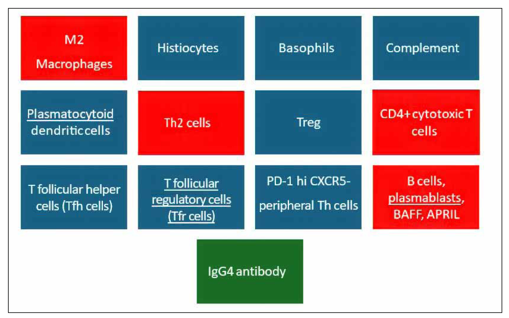
Figure 1. The immune components of the IgG4-related disease.
IgG4-RD requires all the following histopathological findings: a) tissue infiltration by IgG4-positive plasma cells and lymphocytes, b) storiform (whorled) fibrosis patterns, and c) obliterative phlebitis, which is defined as inflammation leading to the occlusion of veins [1-7]. The lymphoplasmacytic infiltrate consists of plasma cells and T cells diffusely distributed in the area of inflammation, while B cells and plasma cells tend to form extranodal germinal centers [1,3]. IgG4-positive plasma cells typically predominate over other types of plasma cells, making up more than 50% of total plasma cells [1]. Notably, the cut-off value for IgG4-positive plasma cells in the visual field is organ-specific and ranges from 10 to 200, depending on the affected organ [1,2]. Polyclonality is a typical finding in the population of lymphocytes, but oligoclonal cases have been reported [1-4]. Tissue eosinophilia is a common histopathological finding in the context of a Th2 response; extensive infiltration is observed in eosinophilic angiocentric fibrosis and eosinophilic cholangitis, resembling allergic disorders and eosinophilic granulomatosis with polyangiitis [1-3,6,7]. Neutrophils and granulomas are atypical findings, although neutrophils are observed in lesions of the respiratory and gastrointestinal mucosa [1,6]. Fibrosis exhibits a patchy distribution with a radial arrangement of collagen fibers, a feature highly specific to IgG4-RD [2,3]. It should be noted that in advanced stages of IgG4-RD, fibrosis predominates over lymphoplasmacytic infiltrate, complicating the diagnosis and management [2,8]. Obliterative phlebitis presents as a lymphoplasmacytic infiltrate in the vascular wall, without necrosis, that occludes the lumen of medium-sized veins; a similar pattern sometimes affects the arteries, especially in the lungs and the pancreas [2,6,7]. The degree of obstruction is variable, but phlebitis without obstruction is also commonly observed [3,7].
The role of IgG4 in the pathogenesis of IgG4-RD is a controversial topic. As mentioned earlier, IgG4 normally causes immunosuppression; thus, its involvement in an immune attack against autoantigens is paradoxical [1-3]. The three main theories claim that a) IgG4 causes direct tissue injury, b) IgG4 has a neutral effect on tissue damage, and c) IgG4 protects the tissue from further damage [1-3]. According to the first theory, the initial inflammatory stimulus triggers the production of IgG4, resulting in the formation of toxic immune complexes [1,3]. According to the second theory, the initial inflammatory stimulus induces both the production of IgG4 and the activation of immune cells that release profibrotic cytokines, which means that IgG4 is a byproduct of this disorder [1,3]. According to the third theory, IgG4 is overproduced as a compensatory mechanism to decrease the activity of the immune disorder [2,3]. The underlying cause of this controversy is the lack of knowledge about the initial inflammatory stimulus; hence, its relationship with the production of IgG4 is unknown [8]. Another cause is the lack of evidence regarding the degree of Fab-arm exchange in patients with IgG4-RD; thus, the ability of pathological IgG4 to form immune complexes, leading to tissue deposition and inflammation, is unclear [2].
A key component of IgG4-RD pathophysiology is the role of CD4+ cytotoxic T lymphocytes (CD4+ CTLs), which are believed to drive tissue inflammation and fibrosis [7] (Figure 2). Unlike in typical immune responses, where CD8+ cytotoxic cells play a significant role, IgG4-RD features an unusual expansion of CD4+ CTLs that express cytotoxic molecules, such as granzyme B and perforin, comprising the majority of the CD4+ infiltrate [9,10]. It is important to note that the presence of CD4+ cytotoxic cells is associated with the accumulation of CD8+ CTLs in the inflammatory area, attributed to the release of self-peptides by the cytotoxic activity of CD4+ CTLs and their presentation on the surface of B cells [9,11]. These cells contribute to chronic tissue damage by inducing apoptosis of resident cells. Consequently, tissue repair is activated, promoting the activation of fibroblasts and enhancing collagen deposition, leading to fibrosis. As a result, the presence of CD4+ CTLs correlates with disease activity, and their decline indicates an adequate response to treatment; however, further investigation is necessary to clarify the association between the levels of CD8+ CTLs and disease activity [8,11].
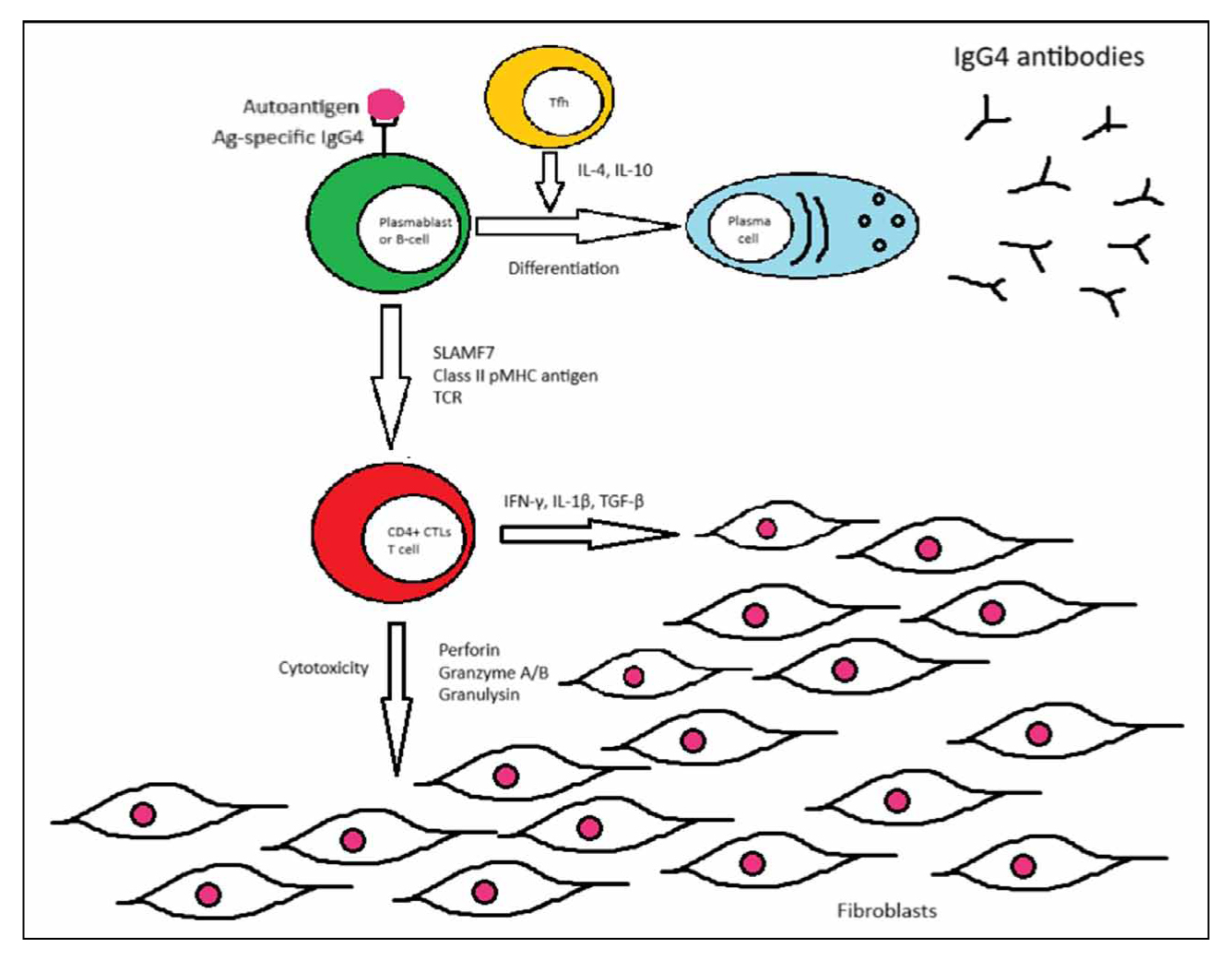
Figure 2. The pathogenesis of IgG4-related disease.
Tregs and activated B cells play a role in disease modulation [8]. Generally, the function of Treg cells is impaired in autoimmune diseases, but this rule does not apply to IgG4-RD [1]. Although this is a controversial topic, increased numbers of Tregs in the peripheral blood and the area of inflammation have been associated with the release of IL-5, IL-10, IL-13, and tumor growth factor-β (TGF-β), leading to increased production of IgG4 and fibrosis, respectively [1,3,8]. In addition, activated Breg cells may contribute to the elevation of IL-10 and TGF-β, and their presence is associated with pancreatic involvement [1,3,4]. Of course, regulatory immune cells possibly represent a compensatory mechanism to control excessive immune activation against normal tissues. B cells are responsible for antigen presentation to CD4+ and perhaps CD8+ T cells and are precursors of IgG4+ plasma cells [2,8,11].
Although the transition from the inflammatory phase to the fibrotic phase is less clear, emerging research has also implicated the role of macrophage and fibroblast interactions in the development of progressive organ fibrosis [8]. Th2-skewed immune responses involving regulatory cells, along with cytokines such as IL-4, IL-5, IL-10, IL-13, interferon-γ (IFN-γ), and TGF-β, are thought to be essential for the activation of eosinophils, alternatively activated macrophages, myofibroblasts, and fibroblasts [3,7,8]. However, the importance of Th2 cells in the pathogenesis of IgG4-RD has been questioned, as they represent a minority of the inflammatory infiltrate, and their elevation is especially linked with atopic disease, so further research is necessary [3,7,8,11,12]. It seems that innate immune cells, such as M2 macrophages, release cytokines that induce the deposition of fibrous tissue [8]. Interestingly, myofibroblasts and fibroblasts are involved in the active phase of the disease, while persistent activation of fibroblasts in the chronic phase leads to excessive collagen production, eventually leading to structural organ damage [3].
Clinicoradiological manifestations and differential diagnosis
IgG4-RD tends to affect middle-aged or elderly male patients; however, it depends on the affected organ and the nationality [1-3,7,8,13-16]. This observation contradicts the general rule that autoimmunity is more common in young female patients [1-3]. For example, significant male predominance has been reported in pancreatic, renal, and retroperitoneal disease, while head and neck involvement is equally distributed between the sexes [2,13,17,18].
IgG4-RD is typically presented as a subacute or chronic disease with nonspecific complaints or as an incidental finding in imaging or histopathological studies [1-3,7,9,19,20]. The clinical findings are usually related to the organs involved; signs and symptoms of acute illness or systemic inflammation are unlikely to occur [1-3,9]. Interestingly, fever is an exclusion criterion for IgG4-RD in contrast to other rheumatic diseases [9,16,19]. However, weight loss might occur, but this is probably a consequence of malabsorption in patients with long-standing pancreatic or biliary involvement [2,3,7,9,19,21]. The indolent course of the disease explains why most patients (~60%) at the time of diagnosis have already developed fibrosis and loss of function, which is partially irreversible [1,22]. The number of affected organs is variable, and it may change over time [1,3]. Most patients present clinically evident illness of a single site, but further investigation commonly reveals asymptomatic involvement of other organs [1,19]. It should be noted that multisystem involvement can occur simultaneously or metachronously [1,3]. In addition, self-limited disease has been reported in a minority of patients [1,3]. Atopic diseases are commonly observed in patients with IgG4-RD, but their relationship is questionable [1-4,7,19,23,24]. Although IgG4-RD can affect any organ, it is categorized into four main clinical patterns based on the organs involved and their epidemiological and pathological features, but there are many exceptions to this rule [4,8,19, 25] (Table 2).
Regarding the abdominal manifestations, IgG4-RD is associated with type 1 autoimmune pancreatitis (type 1 AIP), IgG4-RD sclerosing cholangitis, liver disease, mesenteric involvement, and gastritis. Type 1 AIP presents with focal or diffuse a pancreatic enlargement, causing biliary obstruction, pancreatic insufficiency, and possible involvement of the portal vein or splenic vein [2,3,15,26]. Painless jaundice, weight loss, and diabetes mellitus are common clinical features, while acute pancreatitis is a rare complication [2,3,7,19,26]. Interestingly, endocrine dysfunction with glucagon hypersecretion leading to hyperglycemia has been noted in patients with IgG4-RD without radiologic evidence of pancreatitis [27]. In addition, type 1 AIP has been linked with pancreatic cancer and other malignancies, but further studies are necessary to confirm this association [4,19,28,29]. Furthermore, the causality remains unclear; chronic inflammation triggers cellular proliferation and the development of mutations, but AIP could also represent a paraneoplastic complication of a pre-existing neoplasm [28,29]. Imaging may reveal a sausage-shaped pancreas, a tumor-like lesion, diffuse stenosis of the pancreatic duct, a peripancreatic halo (due to involvement of the peripancreatic fat), pancreatic stones, and pancreatic atrophy (in long-standing disease) [1,2,3,7,26]. On the contrary, pancreatic cancer causes focal stenosis with upstream dilation of the pancreatic duct, while type 2 AIP is more likely to present with an acute episode of pancreatitis, inflammatory bowel disease, and a neutrophilic infiltrate on biopsy [26]. IgG4-RD sclerosing cholangitis is related to type 1 AIP and shares a similar clinical and radiological presentation with primary sclerosing cholangitis (PSC); both diseases present with obstructive jaundice leading to liver cirrhosis and a combination of biliary strictures and dilations on cholangiography [2,3,9,19,30]. However, IgG4-RD is associated with cholecystitis (typically manifesting as subclinical thickening of the gallbladder wall without cholelithiasis) and can also cause pseudo-tumors resembling cholangiocarcinoma [1,2,7,9,19]. Liver involvement should be differentiated from autoimmune hepatitis and liver malignancy due to the abnormal liver function tests and the development of inflammatory pseudotumors [2,3,31]. Inflammation of the mesenteric fat is associated with immobilization of the visceral organs, leading to intestinal obstruction [3,32]. IgG4-related gastritis might be symptomatic, presenting as atrophic mucosa, erosions, ulcers, thickened wall, and submucosal tumors; thus, atrophic gastritis, Helicobacter pylori infection, Castleman disease, and gastric malignancy should be excluded [3,33,34].
IgG4-RD can cause head and neck disease, especially involving the salivary glands, lacrimal glands, orbits, and thyroid gland. Non-tender or slightly tender and typically bilateral involvement of major (and sometimes minor) salivary glands is common [2,7,16,19,30,35,36]. Enlargement of the submandibular glands, known as Kuttner’s tumor, is the most common form of salivary gland involvement and is specific to IgG4-RD [1,3,35]. Mikulicz’s disease involves symmetrical enlargement of the lacrimal, parotid, and submandibular glands [1-3,8-10,30]. It is important to note that sialadenitis caused by IgG4-RD is linked to abnormal saliva flow and the development of sialolithiasis [35]. Dacryoadenitis tends to be bilateral, leading to orbital masses, while dacryocystitis more commonly causes xerophthalmia [3,7,16,30]. Sjögren syndrome is the main differential diagnosis, but IgG4-related salivary and lacrimal gland disease tends to cause mild xerophthalmia or xerostomia that resolves after the administration of corticosteroids. Proptosis, diplopia, and blurry vision may result from swelling of the lacrimal glands or orbital myositis, or both [2,3,7,19,30]. Interestingly, some cases report thickening or compression of nearby nerves [2,3]. Scleritis and uveitis are rare eye-related complications of IgG4-RD [7]. Riedel’s thyroiditis causes fibrosis and progressive, painless enlargement of the thyroid gland (rocky goiter), with reduced blood flow and iodine uptake, leading to tracheal compression, laryngeal nerve palsy, cervical lymphadenopathy, thyroid dysfunction (usually hypothyroidism), and rare involvement of the parathyroid glands [1-3,37,38]. Hence, Hashimoto thyroiditis and thyroid cancer should be excluded based on imaging and histopathology.
IgG4-RD affects the retroperitoneal structures, such as the aorta, kidneys, and ureters. It accounts for a significant portion of idiopathic retroperitoneal fibrosis, which involves connective tissue around major vessels, the upper urinary system, and pelvic structures [1-4,7,16,19,30,39-41]. Common but nonspecific symptoms include back pain or abdominal pain, sometimes radiating to the inguinal areas or thighs [2,40]. However, the overall clinical presentation varies depending on organ-specific complications [39-41]. Moreover, chronic periaortitis (CP) involves inflammatory tissue around the aorta, while chronic aortitis primarily affects the vascular wall [39,40]. Their overlap often makes differentiation difficult; aortitis tends to damage the media, particularly in the thoracic aorta, and can lead to aneurysm formation, whereas periaortitis mainly affects the adventitia and the abdominal aorta [40]. Although the aorta—especially its infrarenal segment extending to the iliac arteries—is most frequently involved, medium-sized branches and retroperitoneal veins can also be affected [2,4,39,40]. Consequently, vascular involvement may manifest as a pulsatile mass, chronic limb ischemia, mesenteric ischemia, coronary artery disease, lower extremity edema, deep venous thrombosis, thrombophlebitis, hydrocele, and varicocele [2,4,39]. The rupture of an inflammatory aneurysm is a rare but life-threatening complication. Vascular imaging often reveals wall thickening (diffuse or localized, sometimes mimicking a mass), dissection, or aneurysms rather than stenosis or strictures [1,3,7,19,30,40]. Fibrous tissue deposition in the retroperitoneal space can irritate or obstruct nearby structures, causing nonspecific symptoms like dysuria and urgency, obstructive uropathy, or renovascular hypertension [39,41]. Regarding the differential diagnosis, rheumatic diseases, such as Takayasu arteritis and giant cell arteritis, tend to affect females and present with fever or arthralgia, while atherosclerosis lacks elevated inflammatory markers [40]. In addition, the exclusion of infectious diseases and malignancies as causes of retroperitoneal fibrosis is recommended [39,40]. Chronic kidney disease can also arise from IgG4-related tubulointerstitial nephritis, leading to proteinuria, hematuria, and potentially progressing to end-stage renal disease [1-3,7,19,30,39,41]. Drug-induced interstitial nephritis, lupus nephritis, and vasculitis should be excluded based on biopsy results [19]. A minority of patients develop nephrotic syndrome due to secondary membranous nephropathy, which differs from primary membranous nephropathy caused by antibodies against phospholipase A2 [2,3,7,30,41]. Renal imaging may reveal enlarged kidneys due to inflammation, atrophic kidneys resulting from long-standing disease, or multiple tumor-like lesions with lower contrast enhancement (with solitary masses being rare), which should be differentiated from renal malignancy [2,3,7,30,39,41].
IgG4-RD can sometimes affect other organs, including the lymph nodes, lungs, serous cavities, skin, nervous system, prostate, and mammary glands (Table 3) [2,3,7,19,30,42-58].
Diagnosis
The diagnosis of IgG4-RD is challenging and depends on the combination of clinical, laboratory, radiological, and histopathological findings. The gold standard is the biopsy result, which should be correlated with the clinical presentation and imaging findings [2,3,30,59]. A significant exception to this rule is type 1 AIP; treatment is indicated in the presence of a typical clinical and radiological presentation without a biopsy [2].
Laboratory testing involves measuring antibodies, complement, inflammatory markers, complete blood count, and plasmablasts/plasma cells. Elevated serum IgG4 (>135 mg/dL) is a useful diagnostic marker, but many patients show normal levels at diagnosis [1-3,7-9,59]. Notably, the upper normal value varies depending on the use of new reagents [60]. Elevated cerebrospinal fluid (CSF) IgG4 suggests immune-mediated inflammation. A recent meta-analysis reports the sensitivity and specificity of IgG4 titers as 87.2% and 82.6%, respectively, though these figures depend on race and disease extent [61].
Adjusting the cutoff value to enhance diagnostic accuracy remains controversial and is currently under investigation [5]. The prozone phenomenon explains the false-negative results in some patients with highly abnormal levels of IgG4; thus, proper serum dilution is recommended to prevent misdiagnosis [2,3,5,8,9]. If the IgG4 levels are normal, a ratio of IgG4/IgG > 10% is suggestive of IgG4-RD, but this remains a controversial marker [2,3,5,7-9]. The nonspecific nature of IgG4 elevation is observed in malignant, autoimmune, and infectious diseases [4]. Other autoantibodies, such as rheumatoid factor, antinuclear antibodies, anti-carbonic anhydrase II antibodies, and anti-smooth muscle antibodies, may also be present [8,15,60,62]. Positive titers of highly specific antibodies for rheumatic diseases are inconsistent with IgG4-RD [16]. Elevation of IgE often occurs with a Th2 response, even without atopy, but this is nonspecific [1,3,4]. Polyclonal gammopathy and elevation of other IgG subclasses, including IgG1 and IgG3, are common; these subclasses activate the complement cascade in many patients [8,15]. Moreover, hypocomplementemia is especially associated with kidney involvement [7,8]. Mild increases in inflammatory markers like C-reactive protein and erythrocyte sedimentation rate mainly involve the aorta and retroperitoneum [7,8]. Eosinophilia is a frequent abnormality in peripheral blood, but highly abnormal counts (>3,000/mm3) as well as leukopenia and thrombocytopenia suggest an alternative diagnosis [8,16]. Plasmablasts, which are intermediate cells between B cells and plasma cells, are released from the bone marrow in response to pathogens, vaccines, and self-antigens. Their elevation has been suggested as a highly sensitive and specific biomarker for IgG4-RD, but further clinical research is needed [2,7,62-65].
Imaging provides crucial diagnostic information after careful assessment of the clinical presentation. The radiological findings are often nonspecific and can be misleading in diagnosing cancer. Different imaging techniques are used based on the involved site. Computed tomography (CT) is the most frequently used test. Magnetic resonance imaging (MRI) is often employed, especially when the biliary tree, the aorta, or the central nervous system is involved [9,40,54]. MRI is an alternative to CT for those with abnormal renal function [41]. Ultrasound is valuable for examining masses in the head and neck region as well as the aorta [40]. Typical findings include organ enlargement, fibrosis, and nodular or mass-like lesions [1,2]. Positron emission tomography (PET) scanning is useful for staging and guiding the site of biopsy due to its higher sensitivity [7,8,66-68]. It should be noted that PET is more likely to recognize arterial, salivary gland, and lymph node pathology, while it lacks sensitivity for renal or brain pathology as well as small, non-active lesions [9,40,68].
Regarding the histopathologic findings, the hallmark of IgG4-RD includes dense lymphoplasmacytic infiltrates, storiform fibrosis, and obliterative phlebitis. Although biopsies from the brain, meninges, pancreas, great vessels, and retroperitoneum are demanding, larger tissue samples are associated with a higher diagnostic yield [2,8]. Sampling from multiple sites or repeat biopsies may be necessary [2]. For example, submandibular gland biopsy is more likely to be diagnostic than labial gland biopsy for IgG4-related salivary gland disease [2]. However, histopathological diagnosis of IgG4-RD is often established after a needle biopsy; endoscopic and surgical biopsies are currently preferred when there is clinical suspicion of IgG4-RD [6]. Furthermore, needle biopsies often fail to recognize the pattern of storiform fibrosis or obliterative phlebitis, while the peripheral infiltrate of IgG4-positive plasma cells in neoplasms might be misdiagnosed as IgG4-RD [6]. Organ-specific interpretation of immunohistochemistry is mandatory, given the variable cut-off value of IgG4-positive plasma cells, ranging from 10 to 200 per high-power field [6]. However, tissue infiltration by IgG4-positive plasma cells is also observed in other diseases of inflammatory and neoplastic etiology; diffuse presence of IgG4-positive plasma cells and a ratio of IgG4 to IgG-positive plasma cells of more than 40% may increase specificity [6,30]. Plasma cells secreting IgE, IgG1, IgG2, and IgG3 represent a minority of the inflammatory infiltrate [3].
Recent advances in the diagnostic field have established general criteria for diagnosing IgG4-RD [30]. These guidelines allow proper diagnosis and treatment even in the absence of typical serological and histopathological findings [30]. Exclusion criteria from the clinical presentation, laboratory measurements, imaging, and biopsy results should be examined during the investigation [30]. Notably, a significant portion of IgG4-RD cases is missed by the general diagnostic criteria in clinical practice due to atypical presentation or involvement of atypical sites [30]. Consequently, organ-specific criteria have been developed to maximize the sensitivity for type 1 AIP, Mikulicz’s disease, and kidney disease [30]. For example, they enable the diagnosis of type 1 AIP based on imaging, making a biopsy unnecessary. In cases where it is difficult to obtain a biopsy, a successful trial of corticosteroids can indirectly confirm the presumptive diagnosis of IgG4-RD [30]. However, physicians should maximize their efforts to obtain a tissue sample for the exclusion of malignancy, especially when the response to corticosteroids is inadequate [8].
Treatment
Fibrosis is a pathological process characterized by excessive tissue scarring, often resulting in organ dysfunction. Preventing fibrosis is crucial for maintaining organ health and avoiding severe complications. Advancements in early diagnosis and appropriate treatment allow successful outcomes. It is vital to start treating before the establishment of fibrosis [1,8]. However, relapses are common, necessitating long-term follow-up. Evidence is still lacking due to insufficient clinical data [8,9].
Treatment of IgG4-RD is indicated under certain circumstances. All patients with symptomatic disease, subclinical involvement of high-risk organs (pancreas, extrapancreatic biliary tree, meninges, aorta, kidney, retroperitoneum, pericardium), subclinical multi-organ disease, or active progression of pre-existing imaging abnormalities are candidates for immunosuppressive treatment [7,19]. The choice of “watch and wait” is appropriate for patients with asymptomatic and stable disease limited to organs with a low risk of severe complications and fibrosis [1,7,19]. Medical treatment is divided into two main phases: induction and maintenance, as in rheumatic diseases [7]. Interventional treatment may be necessary for the management of anatomical abnormalities, such as biliary stenting, ureteral stenting, and nephrostomy tubes [7,19]. Radiation therapy is rarely used as a last-line treatment [9].
Pharmacologic agents for managing IgG4-RD include corticosteroids, non-steroidal alternatives, and biologic agents [1,2,9]. Corticosteroids are the first-line therapy for inducing remission, and they are often used for maintenance [1,2,19]. Disease-modifying antirheumatic drugs (DMARDs), such as azathioprine, mycophenolate mofetil, leflunomide, methotrexate, cyclosporine, iguratimod, and cyclophosphamide, are typically used for maintenance, but the evidence remains limited [1,2]. Recently, various biologic agents have been studied, with rituximab being the most common [1]. Although corticosteroids are considered the cornerstone of treatment in IgG4-RD, most patients will eventually receive non-steroidal immunosuppressants to achieve remission and reduce corticosteroid toxicity [9].
Induction of treatment is the first step in IgG4-RD to decrease the disease’s activity. The drug regimen typically involves corticosteroids; rituximab or methotrexate are alternatives if corticosteroids are strongly contraindicated [1,2,7]. The recommendations about the dose and the duration of the regimen vary depending on the clinical scenario and the expert’s experience. Initially, a medium dose of prednisone, administered orally at 0.4-0.6 mg/kg/day (30-40 mg/day), is administered until resolution, which typically occurs after 2-4 weeks [1,2,7,8,15,19]. Occasionally, a pulse of intravenous methylprednisolone (100-1,000 mg/day for 3 days) is used before the per os regimen for acute complications, such as acute kidney injury or neurological complications [1,7,8]. High doses seem to have similar efficacy to the medium dose of oral corticosteroids [7,8]. Lower doses may be considered when the symptoms are mild. A careful taper should be followed; otherwise, faster withdrawal increases the risk of exacerbation [7,8]. Tapering can be achieved with the following methods: a) reduction of the dose at a rate of 5 mg every two weeks until a final daily dose of 7.5 mg, or b) reduction of the dose at a rate of 10 mg every two weeks until a daily dose of 20 mg, followed a few weeks later with a reduction at a rate of 5 mg every two weeks [1,2]. The total duration of tapering is generally 3-6 months [1,2,7,19].
Maintenance of treatment is achieved with non-steroidal immunosuppressants or low-dose corticosteroids [8]. The addition of nonsteroidal immunosuppressants in the induction phase is indicated in certain patients at increased risk for recurrence (elevated IgE, eosinophilia, elevated IgG4, multiorgan involvement, involvement of high-risk organs, hypocomplementemia) [7,8,9,19]. All patients with a relapse should receive the aforementioned taper of corticosteroids followed by a maintenance plan [7,8]. Japanese experts tend to continue low-dose corticosteroids in the majority of patients for up to three years, but other physicians prefer the combination regimen to discontinue corticosteroids as soon as possible [7]. Rituximab may represent the most effective DMARD, but further research is needed on dosing [1,2,7,8]. Interestingly, the choice of DMARDs varies depending on the organ involved [19].
Regular follow-up, e.g., every six months, with several laboratory tests and imaging techniques, helps assess disease activity and treatment response. However, the management should be guided by these tests after correlation with clinical response. Elevation of serum antibodies (IgG4, IgE, and IgG4/IgG), CSF IgG4, and eosinophils is associated with more active disease [7,8]. It should be noted that changes in the concentration of IgG4 predict relapses in those with elevated IgG4 at baseline [7,8]. Hypocomplementemia occurs during recurrence, implying kidney disease [7,8]. The association of plasmablasts with disease activity is promising, but their use in clinical practice is currently limited [7,8]. Among imaging studies, a PET scan shows active sites of inflammation but cannot differentiate lymphadenopathy from other diseases [7,8].
CONCLUSION
IgG4-related disease is a complex, multisystem disorder that requires a high index of suspicion for diagnosis. Advances in our understanding of its immunopathogenesis and treatment options have significantly improved patient outcomes. Continued research is necessary to further elucidate its mechanisms, refine diagnostic criteria, and optimize management strategies to prevent long-term complications and improve the quality of life for affected individuals.
Future Directions
Ongoing research is focused on identifying novel biomarkers for early detection, understanding the genetic predisposition of IgG4-RD, and developing targeted therapies to improve treatment efficacy while minimizing treatment-related adverse effects. Large-scale clinical trials are needed to evaluate new therapeutic agents and their long-term impact on disease progression.
Conflict of Interest
None to declare.
Declaration of Funding Sources
None to declare.
Author Contributions Statement
Conceptualization: GE. Data Curation: GE, CC. Investigation: GE, CC. Methodology: GE, CC. Project Administration: GE. Supervision: CC. Validation: GE, CC. Writing – Original Draft: GE, CC. Writing – Review Editing: CC. All authors approve the final version of the manuscript.
REFERENCES
- Stone JH, Zen Y, Deshpande V. IgG4-related disease. N Engl J Med. 2012;366(6):539-51.
- Kamisawa T, Zen Y, Pillai S, Stone JH. IgG4-related disease. Lancet. 2015;385(9976):1460-71.
- Mahajan VS, Mattoo H, Deshpande V, Pillai SS, Stone JH. IgG4-related disease. Annu Rev Pathol. 2014;9:315- 47.
- Wallace ZS, Stone JH. An update on IgG4-related disease. Curr Opin Rheumatol. 2015;27(1):83-90.
- Carruthers MN, Khosroshahi A, Augustin T, Deshpande V, Stone JH. The diagnostic utility of serum IgG4 concentrations in IgG4-related disease. Ann Rheum Dis. 2015;74(1):14-8.
- Deshpande V, Zen Y, Chan JK, Yi EE, Sato Y, Yoshino T, et al. Consensus statement on the pathology of IgG4- related disease. Mod Pathol. 2012;25(9):1181-92.
- Pinheiro FAG, Pereira IA, de Souza AWS, Giardini HAM, Cordeiro RA. IgG4-related disease-rare but you should not forget it. Adv Rheumatol. 2024;64(1):35.
- Lanzillotta M, Mancuso G, Della-Torre E. Advances in the diagnosis and management of IgG4 related disease. BMJ. 2020;369:m1067.
- Perugino CA, Stone JH. IgG4-related disease: an update on pathophysiology and implications for clinical care. Nat Rev Rheumatol. 2020;16(12):702-14.
- Mattoo H, Mahajan VS, Maehara T, Deshpande V, Della-Torre E, Wallace ZS, et al. Clonal expansion of CD4(+) cytotoxic T lymphocytes in patients with IgG4-related disease. J Allergy Clin Immunol. 2016;138(3):825-38.
- Perugino CA, Kaneko N, Maehara T, Mattoo H, Kers J, Allard-Chamard H, et al. CD4+ and CD8+ cytotoxic T lymphocytes may induce mesenchymal cell apoptosis in IgG4-related disease. J Allergy Clin Immunol. 2021;147(1):368-82.
- Mattoo H, Della-Torre E, Mahajan VS, Stone JH, Pillai S. Circulating Th2 memory cells in IgG4-related disease are restricted to a defined subset of subjects with atopy. Allergy. 2014;69(3):399-402.
- Kanno A, Nishimori I, Masamune A, Kikuta K, Hirota M, Kuriyama S, et al; Research Committee on Intractable Diseases of Pancreas. Nationwide epidemiological survey of autoimmune pancreatitis in Japan. Pancreas. 2012;41(6):835-9.
- Karim F, Loeffen J, Bramer W, Westenberg L, Verdijk R, van Hagen M, et al. IgG4-related disease: a systematic review of this unrecognized disease in pediatrics. Pediatr Rheumatol Online J. 2016;14(1):18.
- Sato Y, Notohara K, Kojima M, Takata K, Masaki Y, Yoshino T. IgG4-related disease: historical overview and pathology of hematological disorders. Pathol Int. 2010;60(4):247-58.
- Wallace ZS, Naden RP, Chari S, Choi H, Della-Torre E, Dicaire JF, et al; American College of Rheumatology/European League Against Rheumatism IgG4-Related Disease Classification Criteria Working Group. The 2019 American College of Rheumatology/European League Against Rheumatism Classification Criteria for IgG4-Related Disease. Arthritis Rheumatol. 2020;72(1):7-19.
- Raissian Y, Nasr SH, Larsen CP, Colvin RB, Smyrk TC, Takahashi N, et al. Diagnosis of IgG4-related tubulointerstitial nephritis. J Am Soc Nephrol. 2011;22(7):1343-52.
- Zen Y, Onodera M, Inoue D, Kitao A, Matsui O, Nohara T, et al. Retroperitoneal fibrosis: a clinicopathologic study with respect to immunoglobulin G4. Am J Surg Pathol. 2009;33(12):1833-9.
- Della-Torre E, Lanzillotta M, Doglioni C. Immunology of IgG4-related disease. Clin Exp Immunol. 2015;181(2):191-206.
- Stone JH. IgG4-related disease: nomenclature, clinical features, and treatment. Semin Diagn Pathol. 2012;29(4):177-90.
- Herta T, Schröder M, Geisel D, Engelmann C, Tacke F. Management of IgG4-related cholangitis: diagnosis, therapy, and long-term surveillance. Gastroenterol Rep (Oxf). 2025;13:goaf032.
- Wallace ZS, Deshpande V, Mattoo H, Mahajan VS, Kulikova M, Pillai S, et al. IgG4-related disease: clinical and laboratory features in one hundred twenty-five patients. Arthritis Rheumatol. 2015;67(9):2466-75.
- Della Torre E, Mattoo H, Mahajan VS, Carruthers M, Pillai S, Stone JH. Prevalence of atopy, eosinophilia, and IgE elevation in IgG4-related disease. Allergy. 2014;69(2):269-72.
- Inoue D, Yoshida K, Yoneda N, Ozaki K, Matsubara T, Nagai K, et al. IgG4-related disease: dataset of 235 consecutive patients. Medicine (Baltimore). 2015;94(15):e680.
- Wallace ZS, Zhang Y, Perugino CA, Naden R, Choi HK, Stone JH, et al. IgG4-RD Classification Criteria Committee. Clinical phenotypes of IgG4-related disease: an analysis of two international cross-sectional cohorts. Ann Rheum Dis. 2019;78(3):406-12.
- Pastras P, Aggeletopoulou I, Triantos, C. Understanding and management of autoimmune pancreatitis. Achaiki Iatriki. 2025; 44(2):67-70.
- Ito N, Yagi K, Kawano M, Mori Y, Okazaki S, Chujo D, et al. Analysis of pancreatic endocrine function in patients with IgG4-related diseases, in whom autoimmune pancreatitis was ruled out by diagnostic imaging. Endocr J. 2014;61(8):765-72.
- Hart PA, Law RJ, Dierkhising RA, Smyrk TC, Takahashi N, Chari ST. Risk of cancer in autoimmune pancreatitis: a case-control study and review of the literature. Pancreas. 2014;43(3):417-21.
- Schneider A, Hirth M, Münch M, Weiss C, Löhr JM, Ebert MP, et al. Risk of cancer in patients with autoimmune pancreatitis: a single-center experience from Germany. Digestion. 2017;95(2):172-80.
- Umehara H, Okazaki K, Masaki Y, Kawano M, Yamamoto M, Saeki T, et al. Comprehensive diagnostic criteria for IgG4-related disease (IgG4-RD), 2011. Mod Rheumatol. 2012;22(1):21-30.
- Minaga K, Watanabe T, Chung H, Kudo M. Autoimmune hepatitis and IgG4-related disease. World J Gastroenterol. 2019;25(19):2308-14.
- Osama MA, Roy S, Rawat S, Rao S. IgG4 related sclerosing encapsulating peritonitis with cocoon formation: An unusual and undescribed presentation. Indian J Pathol Microbiol. 2022;65(3):683-85.
- Sawada H, Czech T, Silangcruz K, Kozai L, Obeidat A, Wien EA, et al. Clinicopathological characteristics of gastric IgG4-related disease: systematic scoping review. J Gastroenterol Hepatol. 2022;37(10):1865-72.
- Zhang X, Jin X, Guan L, Lin X, Li X, Li Y. IgG4-related disease with gastrointestinal involvement: case reports and literature review. Front Immunol. 2022;13:816830.
- Skillington SA, Ogden MA. IgG4-related disease and the salivary glands: a review of pathophysiology, diagnosis, and management. Otolaryngol Clin North Am. 2021;54(3):497-508.
- Fragoulis GE, Zampeli E, Moutsopoulos HM. IgG4-related sialadenitis and Sjögren’s syndrome. OralDis. 2017;23(2):152-6.
- Czarnywojtek A, Pietrończyk K, Thompson LDR, Triantafyllou A, Florek E, Sawicka-Gutaj N, et al. IgG4-related sclerosing thyroiditis (Riedel-Struma): a review of clinicopathological features and management. Virchows Arch. 2023;483(2):133-144.
- Han X, Zhang P, Li J, Liu Z, Lu H, Luo X, et al. Clinical features and treatment efficacy for IgG4-related thyroiditis. Orphanet J Rare Dis. 2021;16(1):324.
- Razok A, Romero Noboa ME, Sami F, Patolia KN, Tanveer S. IgG4-related disease and isolated retroperitoneal fibrosis: a narrative review. ARP Rheumatol. 2023;2(2):155-7.
- Nikiphorou E, Galloway J, Fragoulis GE. Overview of IgG4-related aortitis and periaortitis. A decade since their first description. Autoimmun Rev. 2020;19(12):102694.
- Kawano M, Yamada K. IgG4-related kidney disease and IgG4-related retroperitoneal fibrosis. Semin Liver Dis. 2016;36(3):283-90.
- Cheuk W, Chan JK. Lymphadenopathy of IgG4-related disease: an underdiagnosed and overdiagnosed entity. Semin Diagn Pathol. 2012;29(4):226-34.
- Cheuk W, Yuen HK, Chu SY, Chiu EK, Lam LK, Chan JK. Lymphadenopathy of IgG4-related sclerosing disease. Am J Surg Pathol. 2008;32(5):671-81.
- Morales AT, Cignarella AG, Jabeen IS, Barkin JS, Mirsaeidi M. An update on IgG4-related lung disease. Eur J Intern Med. 2019;66:18-24.
- Zheng X, Zou W, Zou S, Ye J, Bao Z, Hong J, et al. IgG4-related lung disease: a case report and review of the literature. Altern Ther Health Med. 2023;29(8):320-3.
- Guo Q, Ren Y, Wang Q, Pei H, Jiang S. A case report of IgG4-related respiratory disease with pleural effusion and a literature review. Medicine (Baltimore). 2022;101(30):e29338.
- Tajima M, Nagai R, Hiroi Y. IgG4-related cardiovascular disorders. Int Heart J. 2014;55(4):287-95.
- Tong X, Bai M, Wang W, Han Q, Tian P, Fan H. IgG4-related disease involving polyserous effusions with elevated serum interleukin-6 levels: a case report and literature review. Immunol Res. 2017;65(4):944-50.
- Zhu LP, Khan S, Hui YY, Yang B, Wang SY, Sun KD, et al. IgG4-related disease with ascites: report of a case simulating primary peritoneal papillary serous carcinoma. Dig Dis Sci. 2021;66(5):1751-56.
- Serpa Pinto L, Lamas NJ, Campar A, Ferreira A, Cruz AR. Immunoglobulin G4 related-disease: a rare presentation with secondary hypereosinophilic syndrome and eosinophilic ascites. J Med Cases. 2021;12(3):107-11.
- George V, Tammisetti VS, Surabhi VR, Shanbhogue AK. Chronic fibrosing conditions in abdominal imaging. Radiographics. 2013;33(4):1053-80.
- Chatzigrigoriadis C, Goulioumis A, Sperdouli D, Gyftopoulos K. Embryological, anatomical and clinical considerations on pleuroperitoneal communication. Pleura Peritoneum. 2023;8(3):101-11.
- Tokura Y, Yagi H, Yanaguchi H, Majima Y, Kasuya A, Ito T, et al. IgG4-related skin disease. Br J Dermatol. 2014;171(5):959-67.
- Yang L, Smith RJP, Scolding NJ, Rice CM. IgG4-related disease in the nervous system. Pract Neurol. 2025;25(3):217-27.
- Liu H, Wallace ZS, Harvey L, Crotty R, Wu CL, Deshpande V, et al. Prostate and pancreas involvement are linked in IgG4-related disease. Semin Arthritis Rheum. 2020;50(6):1245-51.
- Chougule A, Bal A, Das A, Singh G. IgG4 related sclerosing mastitis: expanding the morphological spectrum of IgG4 related diseases. Pathology. 2015 ;47(1):27-33.
- Yamada R, Horiguchi S, Yamashita T, Kamisawa T. IgG4-related mastitis, a rare disease, can radiologically and histologically mimic malignancy. BMJ Case Rep. 2016;2016:bcr2016214870.
- Itakura M, Horimoto Y, Ishizuka Y, Onagi H, Hayashi T, Kawamoto T, et al. IgG4-related mastitis characterized by skin thickening of the breast: a case report. Surg Case Rep. 2023;9(1):192.
- Khosroshahi A, Wallace ZS, Crowe JL, Akamizu T, Azumi A, Carruthers MN, et al; Second international symposium on IgG4-related disease. International consensus guidance statement on the management and treatment of IgG4-related disease. Arthritis Rheumatol. 2015;67(7):1688-99.
- Usami Y, Sugano M, Uehara T, Koinuma M, Ishimine N, Kawasaki K, et al. Cut-off values of serum IgG4 among three reagents, including a novel IgG4 reagent: a multicenter study. Sci Rep. 2021;11(1):7280.
- Hao M, Liu M, Fan G, Yang X, Li J. Diagnostic value of serum IgG4 for IgG4-related disease: A PRISMA-compliant systematic review and meta-analysis. Medicine (Baltimore). 2016;95(21):e3785.
- Wallace ZS, Mattoo H, Carruthers M, Mahajan VS, Della Torre E, Lee H, et al. Plasmablasts as a biomarker for IgG4-related disease, independent of serum IgG4 concentrations. Ann Rheum Dis. 2015;74(1):190-5.
- Lanzillotta M, Della-Torre E, Stone JH. Roles of plasmablasts and B Cells in IgG4-related disease: implications for therapy and early treatment outcomes. Curr Top Microbiol Immunol. 2017;401:85-92.
- Mattoo H, Mahajan VS, Della-Torre E, Sekigami Y, Carruthers M, Wallace ZS, et al. De novo oligoclonal expansions of circulating plasmablasts in active and relapsing IgG4-related disease. J Allergy Clin Immunol. 2014;134(3):679-87.
- Koarada S, Tashiro S, Tokuda Y, Ono Y, Sadanaga Y, Suematsu R, et al. Subsets of RP105-negative plasmablasts in IgG4-related disease. Ann Rheum Dis. 2014;73(10):e65.
- Berti A, Della-Torre E, Gallivanone F, Canevari C, Milani R, Lanzillotta M, et al. Quantitative measurement of 18F-FDG PET/CT uptake reflects the expansion of circulating plasmablasts in IgG4-related disease. Rheumatology (Oxford). 2017;56(12):2084-92.
- Tsuji S, Iwamoto N, Horai Y, Fujikawa K, Fujita Y, Fukui S, et al. Comparison of the quantitative measurement of 18F-FDG PET/CT and histopathological findings in IgG4-related disease. Clin Exp Rheumatol. 2021;39(6):1338-44.
- Ebbo M, Grados A, Guedj E, Gobert D, Colavolpe C, Zaidan M, et al. Usefulness of 2-[18F]-fluoro-2-deoxy-D-glucose-positron emission tomography/computed tomography for staging and evaluation of treatment response in IgG4-related disease: a retrospective multicenter study. Arthritis Care Res (Hoboken). 2014;66(1):86-96.