ACHAIKI IATRIKI | 2026; 45(3):135–143
Review
Faethontas Eleftheriadis, Antonios Karanasos, Michail Papafaklis, Anastasios Papoutsoglou, Grigorios Tsigkas, Periklis Davlouros
Department of Cardiology, University of Patras, 26504, Rion-Patras, Greece
Received: 31 Mar 2025; Accepted: 22 Aug 2026
Corresponding author: Assist. Prof. Antonios Karanasos, MD, PhD, 1 Hippokratous Av, 26504, Rion, Patra, Greece. E-mail: akaranasos@upatras.gr, akaranasos@hotmail.com
Keywords: Heart failure, cardiac output, heart disease, atrial fibrillation, pulmonary hypertension

Abstract
Heart failure with preserved ejection fraction (HFpEF) presents an increasingly significant clinical challenge due to its heterogeneous pathophysiology and high morbidity and mortality, alongside a paucity of established disease-modifying therapies. Complex pathomechanisms are implicated in HFpEF, encompassing arterial stiffness, microvascular inflammation, and structural cardiac changes, including left atrial dysfunction and pulmonary hypertension, while diagnosis of HFpEF might also be challenging and relies on clinical assessment, cardiac imaging, and biomarkers, with dedicated scoring systems developed to assess the probability of this condition. Compounding the diagnostic challenges, effective treatment strategies for HFpEF remain limited. Although goals of treatment remain similar to those of heart failure with reduced ejection fraction, aiming at functional improvement, reduction of heart failure hospitalizations, and mortality reductions, treatment options for HFpEF remain rather limited. The current review aims to present a brief synopsis of the mechanisms of HFpEF and diagnostic approaches and focuses on summarizing the current status of HFpEF treatment, including non-pharmacological interventions, management of comorbidities, and pharmacological treatment. Current drug options comprise diuretics for symptom management and SGLT2 inhibitors as the primary disease-modifying agents, while new therapies, such as finerenone and semaglutide, are emerging. Ongoing clinical trials aim to refine personalized treatment approaches, reflecting the need for a precision medicine approach to improve outcomes in this complex syndrome.
INTRODUCTION
Heart failure with preserved ejection fraction (HFpEF) is increasingly acknowledged and diagnosed in clinical practice, partly as a result of an ageing population and a rise in contributory comorbidities, including obesity and diabetes. HFpEF has been recognized as a complex clinical syndrome, accounting for at least half of patients with heart failure. Due to its heterogeneous pathophysiology and epidemiological patterns, HFpEF is often challenging to diagnose and to develop diagnostic and therapeutic modalities. Although cardiovascular mortality is lower in HFpEF than in HF with reduced ejection fraction (HFrEF), hospital readmissions are frequent, and quality of life is poor [1,2]. In this review, we focus on the current and investigational treatment strategies for this complex syndrome.
Pathomechanisms of HFpEF
HFpEF was initially thought to be the result of diastolic dysfunction caused by hypertensive left ventricular (LV) remodeling in isolation, exacerbated by abnormal ventricular-arterial coupling and chronotropic incompetence. These mechanisms were noted originally in HFpEF patients with predominantly hypertensive heart disease, where the primary mechanistic hypotheses centered around hypertension leading to LV hypertrophy with stiff and relatively small left ventricles and, eventually, overt HF [3–5]. Over the past two decades, older adults with isolated hypertensive heart disease have given way to multimorbid patients with obesity, diabetes, and metabolic syndrome as the most common clinical profile in HFpEF. This highlights the fact that the pathomechanisms of HFpEF extend beyond adaptive changes to increased afterload and comprise a variety of factors including systemic inflammation, endothelial dysfunction and altered myocardial energetics.
Arterial stiffness
It is becoming increasingly acknowledged that arterial stiffness plays a significant role in the pathogenesis of HFpEF, impacting prognosis, treatment, and diagnosis. Arterial stiffness, a defining characteristic of vascular aging, increases the afterload on the LV, resulting in diastolic dysfunction, a crucial aspect of HFpEF. The progression of the disease is accelerated by elevated arterial stiffness, which is associated with prevalent cardiovascular risk factors in HFpEF, including obesity, diabetes, and hypertension [3,6].
Microvascular inflammation
Comorbidities in HFpEF lead to a systemic inflammatory state that causes the coronary microvascular endothelium to experience oxidative stress. Cardiomyocytes become stiff and proliferate as a result of decreased PKG activity and decreased cardiac NO bioavailability. This process of myocardial remodeling differs from the one observed in HFrEF, where oxidative stress resulting from ischemia, infection, or toxicity causes cellular death by cardiocyte death and thus promotes cardiac remodeling. The new HFpEF model has significant implications for both diagnosis and treatment [7].
Structural changes in cardiac chambers
The most noticeable abnormalities in HFpEF are impairments in the structure and function of the heart, which affect all four chambers. There are changes in LV relaxation and a decrease in LV compliance, both of which are significant factors in the rise in filling pressures. Atrial fibrillation (AF), which may be regarded as a “biomarker” of underlying LA myopathy and indicative of more advanced HFpEF, is often the result of remodeling and dysfunction of the left atrium (LA) caused by chronic, prolonged, or intermittent elevation of LV filling pressures in HFpEF [8]. It has been demonstrated that deficits in LA strain are the most reliable imaging indicators that differentiate HFpEF from noncardiac causes of dyspnea, and that LA function abnormalities are more often associated with adverse outcomes than LV function abnormalities [9]. While most patients with HFpEF have LV dysfunction before LA dysfunction, some patients may progress to a primary LA cardiomyopathy, which causes lower LV filling and higher LA pressure, thus resulting in inadequate cardiac output augmentation during exertion.
Initially, HFpEF is characterized by minor changes in cardiac remodeling and mild functional abnormalities, with elevated LV filling pressures observed exclusively during exercise. Over time, LA function and remodeling gradually deteriorate, leading to secondary atrial functional MR and increased incidence of paroxysmal AF. This is the stage where symptoms become more prominent and tend to also present during rest. The natural course of the syndrome is characterized by progressive additional deterioration of LA remodeling and dysfunction with development of permanent AF, worsening pulmonary hemodynamics and subsequent pulmonary vascular remodeling with vasoconstriction, and eventual development of RV and RA dysfunction. These changes may also lead to further impairments in cardiac output and increased pericardial restraint, findings associated with advanced HFpEF. The different stages of HFpEF progression are therefore the reason for the varying clinical presentation, and treatments may differ depending on presentation. Progression through these stages is not inevitable and may follow distinct tempos. Comorbidities, inflammation, and endothelial dysfunction can manifest differently among individuals, further increasing heterogeneity.
Regardless of the cause, sustained elevation in LA pressures leads to worsening pulmonary hypertension (PH), especially in patients with more severe LA cardiomyopathy. Roughly 80% of patients with HFpEF have PH, which starts as a passive result of downstream LA pressure elevation. However, in many patients, these passive changes eventually lead to pulmonary vascular disease and consequent combined post- and pre-capillary pulmonary hypertension, characterized by vascular remodeling and vasoconstriction that affects the totality of the pulmonary vasculature leading to deteriorations in exercise capacity, congestion, pulmonary diffusion, and eventually in increased mortality. The more extreme and difficult to handle spectrum of HFpEF is the development of overt clinical right HF due to the PH that will eventually lead to additional symptoms due to systemic congestion and worsening cardiac output.
Atrial Fibrillation
More than half of patients with HFpEF experience AF during their lifetime. AF is independently linked to an increased risk of HF hospitalization and death in HFpEF. Since AF is a reliable sign that HFpEF is probably the cause of HF symptoms like dyspnea, the presence of AF has been incorporated into probability scores for the diagnosis of HFpEF [10]. HFpEF contributes to the rise of AF through structural and functional remodeling of the left atrium (LA). HFpEF leads to larger volumes of LA which is a well-established proarrhythmic substrate associated with atrial fibrosis. This contributes to electrical remodeling, increased atrial refractoriness, and development of AF [11,12].
Coronary Artery Disease (CAD)
Patients with HFpEF frequently have coronary artery disease, which is linked to higher mortality and worsening ventricular function. In patients with CAD, revascularization may be linked to improved outcomes and the maintenance of heart function [13].
Diagnosis of HFpEF
Clinical presentation
Identification of symptoms and signs of HF is the initial step in diagnosing HFpEF. The Framingham Diagnostic Criteria describe the major symptoms and signs indicating the presence of HF. A HF diagnosis requires the presence of two major criteria or one major and two minor criteria. Major criteria include orthopnea or paroxysmal nocturnal dyspnea, jugular venous distension, hepatojugular reflux, rales, S3 gallop rhythm, pulmonary edema, and cardiomegaly. The minor criteria include exertional dyspnea, nighttime cough, ankle edema, tachycardia with a heart rate surpassing 120 beats per minute, hepatomegaly, and pleural effusion. Dyspnea or ankle edema are common first presentations of HFpEF [14].
Cardiac imaging
When evaluating patients who may have HFpEF, cardiac imaging is essential. Transthoracic echocardiography (TTE) is the most useful imaging modality for early diagnosis. In addition to assessment of left ventricular ejection fraction (LVEF), the primary criterion used to distinguish between HFrEF and HFpEF, TTE offers detailed information about the anatomy and physiology of the heart. It detects anomalies in the pericardium, cardiac valves, and myocardium, and the data may be useful in the diagnosis of HFpEF [15]. RV size and function, atrial size, and thorough valve examination are all included in TTE evaluation, which addresses anomalies linked to both anatomy and flow. Importantly, TTE is crucial for the assessment of diastolic function by evaluating LV filling and LA pressures.
Additional non-invasive imaging modalities, besides TTE, may be useful for assessing the structure and function of the heart. For both LV and RV, cardiac MRI provides an accurate and reliably repeatable assessment of heart volumes, mass, and EF. Without using ionizing radiation, cardiac magnetic resonance imaging (MRI) offers remarkable anatomical resolution in all areas of the heart and adjacent structures [16]. It aids in the diagnosis of Fabry disease, cardiac sarcoidosis, cardiac amyloidosis, and hypertrophic cardiomyopathy. Ventricular size, EF, and anomalies of wall motion can also be accurately assessed using ECG-gated cardiac computed tomography (CT). Although radionuclide ventriculography exposes patients to ionizing radiation, it provides extremely repeatable LVEF values. A non-invasive imaging method called positron emission tomography (PET) can reveal details on myocardial fibrosis, inflammation, perfusion, and metabolism [17,18].
In some cases, cardiac catheterization may be necessary to directly measure left ventricular filling pressures and confirm the diagnosis of HFpEF, particularly when non-invasive tests are inconclusive or when there is suspicion of other cardiac conditions. Finally, considering the spectrum of presentations and the fact that several patients may only present with exercise-related symptoms, exercise testing during cardiac catheterization may reveal some additional occult cases of HFpEF.
Biomarkers
Natriuretic peptide assay, including B-type natriuretic peptide (BNP) or N-terminal proB-type natriuretic peptide (NT-proBNP), is commonly used to confirm the presence and severity of HFpEF. Generally, the levels of BNP and NT-proBNP are comparable, rendering either one suitable for clinical use [15,19]. When evaluating dyspnea of potentially cardiac origin, measuring BNP and NT-proBNP levels offers important diagnostic insight, particularly when the cause of dyspnea remains uncertain and physical examination yields inconclusive results. However, it is important to note that BNP levels can be influenced by factors other than heart failure, such as age, renal function, and obesity.
Diagnostic algorithms – scores
To assist in the diagnosis of HFpEF, two primary scoring systems are commonly used, the H2FPEF score and HFA-PEFF score. In the H2FPEF score, several factors are used, including hypertension, age, gender, atrial fibrillation, elevated BNP, and diastolic dysfunction [20]. HFA-PEFF is a more comprehensive tool that includes several clinical, echocardiographic, and biomarker parameters to assess the probability of HFpEF [21]. Both scoring systems help to stratify patients based on their likelihood of having HFpEF, guiding clinicians in the diagnostic process. These scoring systems can aid in the diagnostic evaluation but should always be used in conjunction with clinical judgment and additional diagnostic tests such as echocardiography, imaging, and biomarker analysis [20,21].
Treatment strategies
The major goals of treatment for patients with HFpEF are alleviation of symptoms, improvement of functional status, reduction of the risk of hospitalization for HF, and reduction of the associated mortality. Notably, a higher NYHA class is associated with a lower quality of life in HF patients. This often manifests with an increased risk of HF-related hospital admissions, a clinically meaningful endpoint as it has a well-documented association with higher mortality in patients with HF [22]. Considering the high variety of phenotypes and causes of HFpEF, the approach to treatment needs to be inherently personalized, and tailored to the individual patient’s specific clinical profile (Figure 1).
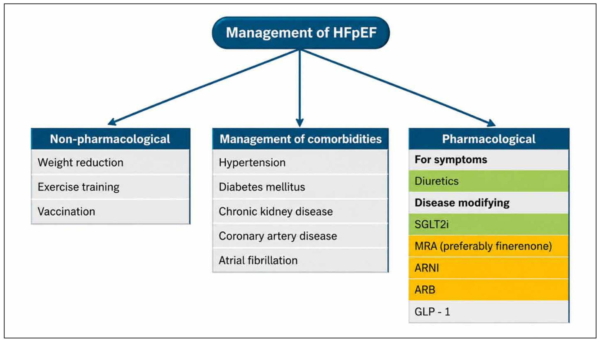
Figure 1. Summary of the treatment of heart failure with preserved ejection fraction. Abbreviations: MRA: mineralocorticoid receptor antagonist; ARNI: angiotensin receptor neprilysin inhibitor; ARB: angiotensin receptor blocker; GLP – 1: glucagon-like peptide 1.
Non-pharmacological treatment
Weight reduction
A higher incidence of heart failure and all-cause death are linked to obesity, particularly central obesity. Up to 80% of people with HFpEF are overweight or obese, and obesity is acknowledged as a significant characteristic of HFpEF [23,24]. Obesity and physical inactivity are also associated with a poor prognosis for HFpEF. About half of the HF risk linked to body mass index (BMI) may be explained by low cardiorespiratory fitness, which has been linked to an increased risk of HF across all BMI categories [25]. Changing one’s lifestyle to reduce weight and improve physical activity are crucial ways to reduce the risk of heart failure.
Exercise training
A significant number of patients with HFpEF have several comorbidities that affect their ability to exercise and contribute to the pathogenesis of HFpEF [26,27]. Exercise-based therapy is becoming a popular non-pharmacological treatment for HFpEF, and it is well known that proper physical activity has many positive impacts on the cardiovascular system. In patients with HFpEF, exercise training enhances peak oxygen consumption and quality of life without significantly altering LV systolic or diastolic function [28].
Vaccination
Pneumonia and respiratory tract infection are important triggers of HF decompensation and cause hospitalization. The risk of pneumonia is high in HF patients especially those with HFpEF [29]. Infections not only increase the in-hospital mortality of HF but also result in poor long-term prognosis [30]. Influenza and pneumococcal vaccines help to prevent respiratory infections that may potentially reduce the risk of exacerbation of HF.
Management of comorbidities
Hypertension and diabetes
Hypertension is the leading cause of HFpEF with a prevalence ranging from 60% to 89% in HFpEF [31]. The systolic blood pressure target for HFpEF should be less than 130 mmHg. The patient’s other comorbidities, such as diabetes, chronic kidney disease (CKD), or coronary artery disease (CAD), should guide the personalized choice of antihypertensive agents. The preferred agents for hypertension control in HFpEF include diuretics, angiotensin receptor blockers (ARB), and mineralocorticoid receptor antagonists (MRA) because these agents also have some beneficial effects for HFpEF, in addition to blood pressure reduction. Since chronotropic incompetence is a potential mechanism contributing to exercise functional limitation in HFpEF, the use of beta-blockers in HFpEF may be avoided if there are no other specific indications for beta-blockers. Given the recently demonstrated benefits of sodium-glucose cotransporter 2 (SGLT2) inhibitors in improving outcomes in patients with HFpEF, SGLT2 inhibitors should be prescribed as first-line therapy for diabetic patients with HFpEF [32]. However, the use of SGLT2 inhibitors is not recommended if the estimated glomerular filtration rate (eGFR) is < 20 mL/min/1.73 m2.
Chronic Kidney Disease
Evidence-based treatments that slow the progression of CKD should be used to treat patients with HFpEF and CKD. It has been demonstrated that SGLT2 inhibitors enhance renal outcomes in CKD patients. After long-term follow-up, the rate of eGFR decline is slower than in patients not on SGLT2 inhibitors, despite an anticipated initial decline in eGFR of about 4 mL/min/1.73 m² upon initiation of SGLT2 inhibitors. Clinical studies also showed that patients receiving angiotensin receptor-neprilysin inhibitors (ARNI) or ARBs experienced a marked slowdown in the fall of eGFR.
Coronary Artery Disease
HFpEF has a high prevalence of CAD. Patients with a low to intermediate pre-test risk of CAD or those with ambiguous non-invasive stress tests should be evaluated for CT coronary angiography in order to assess the severity of CAD and establish the necessity of revascularization. Patients with an intermediate to high pre-test risk of CAD may be candidates for invasive coronary angiography. There are no prospective randomized trials available right now to assess how revascularization affects patients with CAD and HFpEF.
Atrial fibrillation
Anticoagulants continue to be the mainstay of treatment for HFpEF and AF in order to prevent stroke, and the CHA2DS2-VASc score determines when anticoagulation is indicated. There was no evidence in randomized controlled studies that rhythm control with antiarrhythmic drugs was superior to rate control. For rhythm control, ablation of AF might be a more effective method than antiarrhythmic drugs. According to this recommendation, patients who have both HFpEF and AF should have sufficient rate control. First-line medications often consist of beta-blockers and non-dihydropyridine calcium-channel blockers. Digoxin may increase functional capacity and lower NT-proBNP more than bisoprolol in AF patients with heart failure symptoms, according to a recent clinical investigation [33].
Pharmacological treatment
Contrary to HFrEF, where a number of therapies have been demonstrated to improve outcomes including all-cause mortality, HFpEF treatment options have been fairly limited with respect to reducing clinically significant endpoints. Although several treatments with demonstrated benefits in HFrEF have also been evaluated in HFpEF, most of these agents have not been able to demonstrate a consistent benefit in modifying the course of the disease. Therefore, the mainstay of therapy for HFpEF is the use of diuretics for alleviation of congestion symptoms and the use of SGLT2 inhibitors as the only established disease-modifying treatment. It should be noted however, that ongoing research has indicated a potential role for medications such as finerenone and semaglutide in improving outcomes in HFpEF.
Diuretics
HFpEF patients with volume overload should be treated with diuretics [34]. Loop diuretics should be initiated as the first-line therapy with the type and dose depending on the severity of congestion. Loop diuretics (furosemide, torsemide, bumetanide) are typically used, with the dose adjusted to achieve euvolemia (normal fluid status) without causing excessive dehydration or electrolyte imbalances. For those patients with loop diuretic resistance, sequential nephron blockade can be achieved using thiazide diuretics and/or MRAs. Careful monitoring of electrolytes (especially potassium and magnesium) and renal function is essential.
SGLT2 Inhibitors
SGLT2 Inhibitors are the only medication class that has consistently shown improved outcomes in patients with HFpEF. Both empagliflozin and dapagliflozin have shown clear benefits in the management of HFpEF. EMPEROR-Preserved Trial showed that empagliflozin significantly reduced the composite outcome of cardiovascular death or hospitalization for heart failure in patients with HFpEF [32]. Dapagliflozin in the DELIVER Trial also demonstrated a significant reduction in the composite outcome of cardiovascular death or worsening heart failure in HFpEF patients [35]. As SGLT2 inhibitors have received a strong recommendation (Class 2a, Level of Evidence A) in HFpEF guidelines, they should be considered a cornerstone of therapy for all patients with HFpEF, unless contraindicated [36].
Mineralocorticoid Receptor Antagonists (MRAs)
The benefits of spironolactone were observed at the lower end (up to 55%) of HFpEF, however the TOPCAT trial did not show a mortality benefit or a decrease in HF hospitalization throughout the entire study in patients with LVEF 45% or higher. The advantages of candesartan, spironolactone, and ARNI were shown to possibly extend to the lower end of the LVEF range of HFpEF in a meta-analysis conducted at the individual patient level. Thus, MRAs may be taken into consideration to reduce HF hospitalizations in a subset of patients with HFpEF [37]. The precise involvement of MRAs, such as finerenone or spironolactone, in HFpEF is currently being determined by a number of ongoing trials (FINEARTS-HF, SPIRRIT, and SPIRIT-HF) [38]. Recently, results from the FINEARTS-HF trial were published, showing that in patients with HFpEF and HFmrEF, treatment with a non-steroidal MRA, finerenone, was associated with a significantly lower incidence of a composite endpoint consisting of total worsening heart failure events and cardiovascular mortality compared to placebo, giving rise to the possibility for another important disease-modifying therapy in HFpEF [39].
Angiotensin Receptor-Neprilysin Inhibitors (ARNIs)
The PARAGON-HF trial, which evaluated sacubitril/valsartan in HFpEF, did not demonstrate a statistically significant reduction in the primary composite endpoint of total hospitalizations for heart failure and cardiovascular death compared to valsartan alone. However, subgroup analyses suggested a potential benefit in women and in patients with lower ejection fractions (within the preserved range, but closer to 50%). ARNIs may be considered in selected HFpEF patients, especially those with LVEF in the lower range of the preserved spectrum and those who tolerate ACE inhibitors or ARBs well.
Angiotensin Receptor Blockers (ARBs)
Candesartan in the CHARM Preserved trial [40] and irbesartan in the I-Preserve trial [41], failed to achieve a significant reduction in the primary outcome compared with placebo for patients with HFpEF. ARBs can be used in HFpEF patients who are intolerant of ACE inhibitors or ARNIs, primarily for blood pressure control and potential RAAS blockade, but their impact on HFpEF-specific outcomes is limited.
Beta-blockers
In the RATE-AF study that compared digoxin with bisoprolol in AF patients with HF symptoms, the primary outcome of quality of life was comparable between patients treated with bisoprolol and digoxin at 6 months [33]. A beta-blocker is not recommended in patients with HFpEF without compelling indications, such as CAD, prior MI or rate control for AF, especially in those with chronotropic incompetence.
Glucagon-like peptide 1 (GLP-1) agonists– Semaglutide
The STEP-HFpEF-DM and STEP-HFpEF trials showed that semaglutide administration decreased body weight and significantly improved quality of life and the six-minute walk distance in obese patients with HFpEF [42]. There was also a reduction in NT-proBNP levels and notably high reductions in HF-related symptoms and physical limitations in patients with higher NT-proBNP baseline levels. These trials pave the way for a novel disease-modifying therapy in patients with HFpEF, particularly those with a phenotype associated with obesity and cardiometabolic stress.
Future targets
Future therapeutic targets for HFpEF focus on addressing its complex pathophysiology, including inflammation, fibrosis, endothelial dysfunction, and metabolic abnormalities. Key areas of interest include anti-inflammatory and anti-fibrotic agents, enhancement of the NO–cGMP–PKG pathway, and modulation of myocardial energetics and titin stiffness. Other emerging approaches target mitochondrial dysfunction, autonomic imbalance, and coronary microvascular dysfunction. Personalized, phenotype-driven therapies and ongoing clinical trials aim to improve outcomes by defining specific HFpEF phenotypes thus tailoring individualized treatment to the diverse HFpEF population.
A considerable number of clinical trials are currently in progress to explore potential therapeutic options for HFpEF. Given the substantial morbidity and mortality associated with HFpEF, it is imperative that these therapies be more routinely considered and offered to patients as viable adjunctive treatment options. The SUMMIT trial is investigating Tirzepatide in patients with NYHA functional class II-IV, Elevated NT-proBNP, BMI ≥30 kg/m2, CAMEO-SEMA is evaluating semaglutide, SPIRRIT and SPIRIT-HF are investigating Spironolactone vs placebo, and CADENCE is evaluating Sotatercept in patients with EF ≥50% with combined pre- and postcapillary PH PVR ≥4 WU, PCWP >15 but <30 mm Hg are some of the ongoing trials [43] ENDEAVOR is a phase 2b-3 study of Mitiperstat, therapy that targets comorbidity-induced systemic inflammation which may be a key mechanism underlying HFpEF [44] HERMES is evaluating patients with HFpEF and HFmrEF, investigating Ziltivekimab, a monoclonal antibody targeting interleukin-6 to reduce systemic inflammation [45].
CONCLUSIONS
More than half of all HF cases are HFpEF, a growing public health concern characterized by increased morbidity and mortality. HFpEF frequently goes undiagnosed, which results in high health resource usage. Considering the variety of the presenting phenotypes and the lack of established disease-modifying therapies, as in HFrEF, there are considerable challenges in the management of HFpEF, and an unmet clinical need. Currently, research is focused on assessing tailored treatment for different subtypes of HFpEF and thoroughly phenotyping HFpEF patients. Future clinical trials using a precision medicine approach in a variety of HFpEF characteristics might be an important step forward in the attempt toward establishing new pathways for diagnosis and management of HFpEF, thus improving outcomes.
Conflict of interest
The authors declare that the research was conducted in the absence of any commercial or financial relationships that could be construed as a potential conflict of interest.
Declaration of funding sources
AK and MP received funding by the funding program “MEDICUS” of the University of Patras.
Author Contributions statement
Conception and design: FE, AK and PD; analysis and interpretation of the data: FE, AK and AP; drafting of the article: FE, AK and AP; critical revision of the article for important intellectual content: AK, GT, MP and PD; final approval of the article: All authors.
REFERENCES
- Shah KS, Xu H, Matsouaka RA, Bhatt DL, Heidenreich PA, Hernandez AF, et al. Heart Failure With Preserved, Borderline, and Reduced Ejection Fraction: 5-Year Outcomes. J Am Coll Cardiol. 2017; 70(20): 2476–86.
- Reddy YNV, Rikhi A, Obokata M, Shah SJ, Lewis GD, AbouEzzedine OF, et al. Quality of life in heart failure with preserved ejection fraction: importance of obesity, functional capacity, and physical inactivity. Eur J Heart Fail. 2020; 22(6):1009–18.
- Zile MR, Baicu CF, Gaasch WH. Diastolic heart failure-abnormalities in active relaxation and passive stiffness of the left ventricle. N Engl J Med. 2004;350(19):1953–9.
- Borlaug BA, Melenovsky V, Russell SD, Kessler K, Pacak K, Becker LC, et al. Impaired chronotropic and vasodilator reserves limit exercise capacity in patients with heart failure and a preserved ejection fraction. Circulation. 2006; 114(20):2138–47.
- Kawaguchi M, Hay I, Fetics B, Kass DA. Combined ventricular systolic and arterial stiffening in patients with heart failure and preserved ejection fraction: implications for systolic and diastolic reserve limitations. Circulation. 2003; 107(5):714–20.
- Lam CSP, Roger VL, Rodeheffer RJ, Bursi F, Borlaug BA, Ommen SR, et al. Cardiac structure and ventricular-vascular function in persons with heart failure and preserved ejection fraction from Olmsted County, Minnesota. Circulation. 2007; 115(15):1982–90.
- Paulus WJ, Tschöpe C. A novel paradigm for heart failure with preserved ejection fraction: comorbidities drive myocardial dysfunction and remodeling through coronary microvascular endothelial inflammation. J Am Coll Cardiol. 2013;62(4):263–71.
- Melenovsky V, Hwang SJ, Redfield MM, Zakeri R, Lin G, Borlaug BA. Left atrial remodeling and function in advanced heart failure with preserved or reduced ejection fraction. Circ Heart Fail. 2015; 8(2):295–303.
- Telles F, Nanayakkara S, Evans S, Patel HC, Mariani JA, Vizi D, et al. Impaired left atrial strain predicts abnormal exercise haemodynamics in heart failure with preserved ejection fraction. Eur J Heart Fail. 2019; 21(4):495–505.
- von Haehling S, Assmus B, Bekfani T, Dworatzek E, Edelmann F, Hashemi D, et al. Heart failure with preserved ejection fraction: diagnosis, risk assessment, and treatment. Clin Res Cardiol. 2024;113(9):1287-305.
- Knackstedt C, Gramley F, Schimpf T, Mischke K, Zarse M, Plisiene J, et al. Association of echocardiographic atrial size and atrial fibrosis in a sequential model of congestive heart failure and atrial fibrillation. Cardiovasc Pathol. 2008; 17(5): 318–24.
- Karanasos A, Tyrovolas K, Tsiachris D, Efremidis M, Kordalis A, Karmpalioti M et al. Left Atrial Function Post Radiofrequency and Cryoballoon Ablation Assessed by Volume-Pressure Loops. Front Cardiovasc Med. 2022; 9:830055.
- Hwang SJ, Melenovsky V, Borlaug BA. Implications of coronary artery disease in heart failure with preserved ejection fraction. J Am Coll Cardiol. 2014; 63(25 Pt A):2817–27.
- McKee PA, Castelli WP, McNamara PM, Kannel WB. The natural history of congestive heart failure: the Framingham study. N Engl J Med. 1971; 285(26):1441–6.
- Nauta JF, Hummel YM, van der Meer P, Lam CSP, Voors AA, van Melle JP. Correlation with invasive left ventricular filling pressures and prognostic relevance of the echocardiographic diastolic parameters used in the 2016 ESC heart failure guidelines and in the 2016 ASE/EACVI recommendations: a systematic review in patients with heart failure with preserved ejection fraction. Eur J Heart Fai.l 2018; 20(9):1303–11.
- He J, Yang W, Wu W, Yin G, Zhuang B, Xu J, et al. Heart Failure with Normal Natriuretic Peptide Levels and Preserved Ejection Fraction: A Prospective Clinical and Cardiac MRI Study. Radiol Cardiothorac Imaging. 2024; 6(3): e230281.
- Ashkir Z, Myerson S, Neubauer S, Carlhäll CJ, Ebbers T, Raman B. Four-dimensional flow cardiac magnetic resonance assessment of left ventricular diastolic function. Front Cardiovasc Med. 2022; 9:866131.
- Quarta G, Gori M, Iorio A, D’Elia E, Moon JC, Iacovoni A, et al. Cardiac magnetic resonance in heart failure with preserved ejection fraction: myocyte, interstitium, microvascular, and metabolic abnormalities. Eur J Heart Fail. 2020; 22(7):1065–75.
- Booth RA, Hill SA, Don-Wauchope A, Santaguida PL, Oremus M, McKelvie R, et al. Performance of BNP and NT-proBNP for diagnosis of heart failure in primary care patients: a systematic review. Heart Fail Rev. 2014; 19(4):439–51.
- Koch DG, Rockey DC, Litwin SS, Tedford RJ. H2FPEF Scores Are Increased in Patients with NASH Cirrhosis and Are Associated with Post-liver Transplant Heart Failure. Dig Dis Sci. 2024; 69(8): 3061–8.
- Li X, Liang Y, Lin X. Diagnostic and prognostic value of the HFA-PEFF score for heart failure with preserved ejection fraction: a systematic review and meta-analysis. Front Cardiovasc Med. 2024; 11:1389813.
- Iqbal J, Francis L, Reid J, Murray S, Denvir M. Quality of life in patients with chronic heart failure and their carers: a 3-year follow-up study assessing hospitalization and mortality. Eur J Heart Fail. 2010;12(9):1002–8.
- Clerico A, Zaninotto M, Passino C, Plebani M. Obese phenotype and natriuretic peptides in patients with heart failure with preserved ejection fraction. Clin Chem Lab Med. 2018; 56(7):1015–25.
- Kittleson MM, Panjrath GS, Amancherla K, Davis LL, Deswal A, Dixon DL, et al. 2023 ACC Expert Consensus Decision Pathway on Management of Heart Failure With Preserved Ejection Fraction: A Report of the American College of Cardiology Solution Set Oversight Committee. J Am Coll Cardiol. 2023; 81(18):1835–78.
- Pandey A, Cornwell WK, Willis B, Neeland IJ, Gao A, Leonard D, et al. Body Mass Index and Cardiorespiratory Fitness in Mid-Life and Risk of Heart Failure Hospitalization in Older Age: Findings From the Cooper Center Longitudinal Study. JACC Heart Fail. 2017; 5(5):367–74.
- Shah SJ, Kitzman DW, Borlaug BA, Van Heerebeek L, Zile MR, Kass DA, et al. Phenotype-Specific Treatment of Heart Failure With Preserved Ejection Fraction: A Multiorgan Roadmap. Circulation. 2016; 134(1):73–90.
- Sharma K, Kass DA. Heart failure with preserved ejection fraction: mechanisms, clinical features, and therapies. Circ Res. 2014; 115(1): 79–96.
- Pandey A, Parashar A, Kumbhani DJ, Agarwal S, Garg J, Kitzman D, et al. Exercise training in patients with heart failure and preserved ejection fraction: meta-analysis of randomized control trials. Circ Heart Fail. 2015; 8(1):33–40.
- Shen L, Jhund PS, Anand IS, Bhatt AS, Desai AS, Maggioni AP, et al. Incidence and Outcomes of Pneumonia in Patients With Heart Failure. J Am Coll Cardiol. 2021; 77(16):1961–73.
- Chen CY, Lee CH, Lin HW, Lin SH, Li YH. Impact of infection-related admission in patients with heart failure: a 10 years national cohort study. Sci Rep. 2023; 13(1):6941.
- Owan TE, Hodge DO, Herges RM, Jacobsen SJ, Roger VL, Redfield MM. Trends in prevalence and outcome of heart failure with preserved ejection fraction. N Engl J Med. 2006; 355(3): 251–9.
- Anker SD, Butler J, Filippatos G, Ferreira JP, Bocchi E, Böhm M, et al. Empagliflozin in Heart Failure with a Preserved Ejection Fraction. N Engl J Med. 2021; 385(16):1451–61.
- Kotecha D, Bunting K V., Gill SK, Mehta S, Stanbury M, Jones JC, et al. Effect of Digoxin vs Bisoprolol for Heart Rate Control in Atrial Fibrillation on Patient-Reported Quality of Life: The RATE-AF Randomized Clinical Trial. JAMA. 2020;324(24):2497–508.
- McDonagh TA, Metra M, Adamo M, Gardner RS, Baumbach A, Böhm M, et al. 2023 Focused Update of the 2021 ESC Guidelines for the diagnosis and treatment of acute and chronic heart failure. Eur Heart J. 2023; 44(37):3627–39.
- Solomon SD, McMurray JJV, Claggett B, de Boer RA, DeMets D, Hernandez AF, et al. Dapagliflozin in Heart Failure with Mildly Reduced or Preserved Ejection Fraction. N Engl J Med. 2022; 387(12):1089–98.
- Heidenreich PA, Bozkurt B, Aguilar D, Allen LA, Byun JJ, Colvin MM, et al. 2022 AHA/ACC/HFSA Guideline for the Management of Heart Failure: A Report of the American College of Cardiology/American Heart Association Joint Committee on Clinical Practice Guidelines. J Am Coll Cardiol. 2022; 79(17):e263–e421.
- Dewan P, Jackson A, Lam CSP, Pfeffer MA, Zannad F, Pitt B, et al. Interactions between left ventricular ejection fraction, sex and effect of neurohumoral modulators in heart failure. Eur J Heart Fail. 2020; 22(5):898–901.
- Vaduganathan M, Claggett BL, Lam CSP, Pitt B, Senni M, Shah SJ, et al. Finerenone in patients with heart failure with mildly reduced or preserved ejection fraction: Rationale and design of the FINEARTS-HF trial. Eur J Heart Fail. 2024; 26(6): 1324–33.
- Solomon SD, McMurray JJV, Vaduganathan M, Claggett B, Jhund PS, Desai AS, et al. Finerenone in Heart Failure with Mildly Reduced or Preserved Ejection Fraction. N Engl J Med. 2024; 391(16):1475–85.
- Yusuf S, Pfeffer MA, Swedberg K, Granger CB, Held P, McMurray JJV, et al. Effects of candesartan in patients with chronic heart failure and preserved left-ventricular ejection fraction: the CHARM-Preserved Trial. Lancet. 2003; 362(9386):777–81.
- Massie BM, Carson PE, McMurray JJ, Komajda M, McKelvie R, Zile MR, et al. Irbesartan in patients with heart failure and preserved ejection fraction. N Engl J Med, 2008; 359(23):2456–67.
- Kosiborod MN, Abildstrøm SZ, Borlaug BA, Butler J, Rasmussen S, Davies M, et al. Semaglutide in Patients with Heart Failure with Preserved Ejection Fraction and Obesity. N Engl J Med. 2023; 389(12):1069–84.
- Borlaug BA, Sharma K, Shah SJ, Ho JE. Heart Failure With Preserved Ejection Fraction: JACC Scientific Statement. J Am Coll Cardiol. 2023; 81(18): 1810–34.
- Lund LH, Lam CSP, Pizzato PE, Gabrielsen A, Michaëlsson E, Nelander K, et al. Rationale and design of ENDEAVOR: A sequential phase 2b–3 randomized clinical trial to evaluate the effect of myeloperoxidase inhibition on symptoms and exercise capacity in heart failure with preserved or mildly reduced ejection fraction. Eur J Heart Fail. 2023; 25(9):1696–707.
- Ministrini S, Liberale L, Puspitasari YM, Han J, Kirmes K, Unkelbach LP, et al. Direct Interleukin-6 Inhibition Blunts Arterial Thrombosis by Reducing Collagen-Mediated Platelet Activation. Arterioscler Thromb Vasc Biol. 2025; 45(8):1432-9.