ACHAIKI IATRIKI | 2026; 45(3):178–186
Review
Dimitrios Efthymiou1, Iwanna Marlafeka1, Prokopis Karidis1, Maria Lagadinou1,2
1Department of Internal Medicine, University Hospital of Patras, 26504 Patras, Greece
2School of Medicine, University of Patras, 26504 Patras, Greece
Received: 28 Feb 2026; Accepted: 08 Jun 2026
Corresponding author: Dimitrios Efthymiou, MD, PhD(c), Internal Medicine Resident, Department of Internal Medicine, University of Patras, 26504, Rio, Patras, Greece. E-mail: efthdim2@gmail.com
Keywords: Pleural effusion, chylothorax, pleural fluid, pseudochylothorax

Abstract
Pleural effusion is defined as the abnormal accumulation of fluid within the pleural space and represents a common clinical manifestation of diverse cardiopulmonary, infectious, inflammatory, systemic, and malignant disorders. Its development reflects an imbalance between pleural fluid production and reabsorption.
Pleura Anatomy – Innervation – Pleural fluid – Pleural effusion
Lungs are separated from the chest wall by a continuous fibrous mesothelial membrane (serous membrane) that is divided into two layers, parietal pleura and visceral pleura, with the hilum being the transition point. The visceral pleura overlies the lung surface, and the parietal pleura overlies the chest wall. The parietal pleura is further divided into cervical, costal, mediastinal and peripheral pleura based on the underlying structures. This subdivision of the parietal pleura is of utmost importance since each part has distinct somatic innervation, leading to radiation of pain to different neighbouring structures of the same somatic innervation. More specifically, the cervical pleura is innervated by the first thoracic spinal nerve, the costal pleura by the overlying thoracic nerves, the mediastinal pleura by the phrenic nerve, and the peripheral diaphragmatic pleura by the lower six thoracic nerves. On the other hand, the visceral pleura adheres to the lungs and is only innervated by autonomic fibres leading to an absence of pain sensation [1].
The space between the two layers of the pleura is filled with a small amount of fluid, approximately 0.2-0.3 ml/kg per pleural cavity (10-20 ml for a 70kg human per pleural space). The presence of a small amount of pleural fluid plays a crucial role in immune surveillance and facilitates movement between the two layers of the pleura during respiration acting as a natural lubricant. It is continuously produced at a rate of 0.01ml/kg/h, by the systemic circulation, arising from the thoracic pleura into the pleural space with a net driving force of +5 cm H2O. This driving force is the result of the difference between the oncotic and hydrostatic pressures of the systemic and pulmonary circulations. Drainage of the pleural fluid is managed through the lymphatics of parietal pleura which end up draining into the right thoracic or the left thoracic duct. Drainage rate can reach a maximum of 0.3 to 0.4 ml/kg/h [1–3]. More specifically, the thoracic duct is responsible for the lymphatic drainage of the entire body except for the right sides of the head and neck, the right side of the thorax and pleural space, and the right upper extremity, which are drained by the right lymphatic duct [4].
As far as the composition of pleural fluid is concerned it generally contains a normal number of cells, approximately 1.7 x10³ white blood cells per microliter, of which 75% are macrophages, 23% lymphocytes, 1% neutrophils (slightly higher in smokers), 0% eosinophils and 1% to 2% mesothelial cells. Moreover, it contains a small amount of protein, which is less than 1.5 g/dl and is primarily composed of albumin [1,5].
MATERIALS AND METHODS
This narrative review was conducted following a PubMed search using the keywords “pleural effusion,” “pleural fluid,” “pseudochylothorax” and “chylothorax,” limited to English-language publications through February 2026. Twenty-nine relevant articles, including clinical studies, reviews, and guideline-based publications, were selected for analysis.
Mechanisms of Fluid Accumulation
Pleural effusion is the abnormal accumulation of pleural fluid due to local or systemic pathologies that lead to changes in the dynamic balance and equilibrium within the chest cavity [1]. This pathological fluid accumulation is generally the result of an imbalance between the rate of production and the rate of reabsorption of pleural fluid. The underlying pathology affecting the production of fluid may be either systemic or local, and it affects the oncotic and/or hydrostatic pressure or the integrity of the pleura (Table 1). As far as disruption of drainage is concerned, the underlying pathologic mechanisms affect the removal of fluid and mainly disrupt the flow of the lymphatic vessels or lead to increased central venous pressure, which indirectly leads to lymphatic fluid accumulation [2].
Symptoms – Signs – Imaging modalities
Pleural effusion is characterized by a wide range of signs and symptoms varying from asymptomatic disease to exertional breathlessness or dyspnoea and orthopnoea at rest and moderate to profound hypoxemia and tachypnoea with the underlying pathophysiology being vague [6].
Based on the PLEASE study, one of the largest prospective studies regarding pleural effusion signs and symptoms pre- and post- drainage, reducing the space-occupying or mass effect of pleural effusion appears to be an important contributor to improved breathlessness irrespective of lung re-expansion. Notably, in the PLEASE study, lung volumes, heart rate and respiratory rate underwent only modest absolute changes after drainage, whereas the ipsilateral diaphragm showed more obvious changes in shape and movement, possibly due to its more compressible/distortable nature. Abnormalities in diaphragm movement were common, and their presence, in addition to baseline level of breathlessness, were independent predictors of the likelihood of symptom relief after fluid drainage [7]. Another smaller study by Estenne et al. suggested that relief of dyspnoea following thoracocentesis results primarily from a reduction in the size of the thoracic cage, which allows the inspiratory muscles to operate on a more advantageous portion of their length-tension curve. This is because only small and inconsistent changes in pulmonary mechanics were observed and could not explain the immediate and remarkable relief of dyspnoea noted by the patients. In contrast, an invariable shift of the minimal (inspiratory) pleural pressure -volume curve resulted in generated pressures by the inspiratory muscles being markedly more negative at any comparable lung volume [8].
Moreover, symptoms like cough, fever, and systemic signs also depend on the underlying cause of the effusion. Rare though it may be, effusions can attain sizes significant enough to induce hemodynamic changes, imitating tamponade. Of note, patients suffering from pleural inflammation called pleurisy may complain of sharp, severe, localized crescendo-decrescendo pain with inhalation and exhalation or while coughing. However, the characteristic pleuritic pain is expected to subside when pleural effusion develops while constant pain is a hallmark of malignant diseases such as mesothelioma that may not subside even after pleural effusion accumulation [2,9].
Physical examination findings like intercostal space fullness and percussion dullness are appreciated in large effusions. Auscultation reveals decreased breath sounds and absence of tactile and vocal fremitus over the area of the effusion. Before pleural effusion formation, pleural rubs may be auscultated and are often mistaken for coarse crackles, indicating active pleurisy. Based on a systematic review by Camilla L Wong et al., among the different examination maneuvers of conventional percussion, auscultatory percussion, breath sounds, chest expansion, tactile vocal fremitus, vocal resonance, crackles, and pleural friction rub, conventional percussion was most accurate for diagnosing pleural effusion (summary positive likelihood ratio, 8.7; 95% confidence interval, 2.2-33.8), while the absence of reduced tactile vocal fremitus made pleural effusion less likely (negative likelihood ratio, 0.21; 95% confidence interval, 0.12-0.37) [2,9].
Regarding the imaging modalities for pleural effusion detection, chest radiography (CXR), computed tomography (CT) and ultrasound (US) are the most common and useful options. More specifically, standing lateral view chest X-ray can detect small effusions of 50 ml, while standing posterior – anterior view (PA) chest X-ray can detect effusions of 200-300 ml. In both cases, guiding points are vague costophrenic angles. Lateral decubitus chest X- ray is regarded as the most useful X-ray in detecting pleural effusions even as low as 15-50 ml with a sensitivity of 96 % and a specificity of 99% [10–12]. Based on a recent systematic review point of care ultrasound (POCUS) seems to have a higher diagnostic accuracy in identification of pleural effusion (as small as 20ml) as a screening tool compared to radiography with a sensitivity of 94% and a specificity of 97 % even in the hands of inexperienced users. It can also be superior to CT in cases of septated effusions identifying the multiple distinct, walled-off compartments or pockets and even guiding thoracocentesis [11,13].
Finally, regarding the use of CT scan, it can detect effusions lower than 15 ml, identify a possible empyema or a septated effusion and also characterize nearby structures such as the lung parenchyma and lymph nodes. In that way it helps differential diagnosis, clarifying the possible underlying cause and describing the extent of it. A 2024 retrospective analysis assessed the diagnostic accuracy of CT in differentiation of transudate from exudate pleural effusions based on pleural effusion Hounsfield Units (HU) and pleural fluid to blood HU ratio. It showed that exudates had significantly higher CT values and pleural fluid to blood CT ratio than those with transudates. More specifically with a cut-off value of 10.81 HU, the area under the curve (AUC), sensitivity, specificity, positive predictive value (PPV) and negative predictive value (NPV) of CT value were 0.85, 88.89%, 68.90%, 43.96%, and 95.76%, respectively. The optimum cut-off value for PF to blood CT value ratio was 0.27 with AUC of 0.86, yielding a sensitivity of 61.11%, specificity of 86.36%, PPV of 78.57%, and NPV of 73.08%. As a result, CT value and PF to blood CT value ratio showed good differential abilities in predicting transudates from exudates, which may help to avoid unnecessary thoracocentesis [14].
Thoracocentesis
To alleviate symptoms (therapeutic thoracocentesis), differentiate exudate from transudate effusions and guide therapeutic decisions, fluid can be drained from the chest cavity via the minimally invasive technique of thoracocentesis. In this procedure, a 20g needle is inserted in the intercostal space on the upper border of the lower rib to avoid the intercostal neurovascular bundle, typically between the sixth and eighth intercostal space in the midaxillary line if the patient is supine or the posterior mid-scapular line if the patient is seated. In cases where US guidance is used (ideally), the insertion point is usually 1-2 intercostal spaces lower than the upper limit of the effusion but not lower than the 9th rib. When performed with appropriate technique and precautions, it carries a low risk of complications like pneumothorax (5-10%), hemothorax (1%) re-expansion pulmonary oedema (in therapeutic thoracocentesis where >1.5lt of fluid is drained) and spleen or liver laceration. As a result, post procedure CXR is not routinely indicated [3,13,15].
Contraindications for thoracocentesis are coagulation disorders, specifically a platelet count <50.000 /μL or INR > 2 and local active dermal infection or cellulitis. Intubated patients are a relevant contraindication where thoracocentesis should only be performed when pleural fluid thickness is >15mm in >3 consecutive intercostal spaces [15].
Indications for thoracocentesis are the presence of a newly diagnosed pleural effusion larger than 1cm in the CXR decubitus view and over 1cm thickness in the US. However, small bilateral effusions in patients with decompensated heart failure, cirrhosis, or kidney failure with no underlying fever, chest pain or effusion asymmetry, are likely transudative and do not require diagnostic thoracentesis. In these cases, diuresis for up to 48h can be performed leading to resolution of up to 75% of these effusions. In cases where the effusion does not resolve, a diagnostic thoracocentesis is prompted [3,16].
Moreover, parapneumonic effusions occurring as a complication of bacterial pneumonia, lung abscess, or bronchiectasis should be tapped as soon as possible to distinguish complicated from uncomplicated ones and to guide therapeutic decisions. More specifically, complicated parapneumonic effusions are characterized by positive Gram stain or positive culture or pH of pleural fluid < 7.2 or glucose of pleural fluid less than 60 mg/dl orLDH> 1000 IU/L and are in need of drainage with thoracostomy for resolution and not only antibiotic therapy. A special subcategory of complicated parapneumonic effusions is empyema, also known as pyothorax or purulent pleuritis, and it is characterized by a pleural effusion with the gross appearance of frank pus that also needs tube thoracostomy in addition to antibiotic therapy. Also, uncomplicated parapneumonic effusions that exceed 1/2 of the hemithorax should also be managed with tube thoracostomy to avoid organization [3,17,18].
Therapeutic thoracocentesis should also be performed in cases of haemothorax drainage, for symptomatic (dyspnoea, hypoxemia) pleural effusions of more than 300 ml and pre-pleurodesis. In all cases of tube thoracostomy, one should always avoid immediate drainage of >1.5lt of fluid because of the possibility of re-expansion pulmonary edema which is characterised by the early sign of persistent cough post drainage [15].
Pleural fluid work up
In cases of diagnostic thoracocentesis at least 20-30 ml of pleural fluid should be drained and sent to the laboratory. The analysis of pleural aspirate should routinely include a Gram stain, differential cell count, culture, cytology (at least 60ml), protein levels, l-lactate dehydrogenase, and pH. The appearance of the fluid (bloody, clear, straw, turbid, purulent, milky) is also helpful in the diagnostic algorithm. Additional tests, such as tuberculosis (TB) screening with Ziehl-Neelsen stain and culture for common bacteria, Mycobacterium Tuberculosis and nontuberculous (NTM) mycobacteria, should be considered based on individual patient risk factors and regional prevalence [15,19].
Special pleural fluid values to keep in mind are:
- A ratio of pleural fluid Hct / blood Hct >50 %: Indicates hemothorax.
- NT pro-BNP of pleural fluid ≥1500 pg/ml has 91% Se and 93% Sp for congestive heart failure [20]
- ADA of pleural fluid: <40 exclude TB, >70 suggest TB
- Pleural fluid glucose <60mg/dl or pleural fluid glucose/ serum glucose < 0,5: Malignancy, TB pleuritis, Rheumatoid arthritis, Infection, Systemic Lupus Erythematosus
- High Pleural fluid amylase: Indicative of pancreatic disease or esophageal rupture
- Triglycerides >110mg/dL indicate chylothorax
- Pleural fluid cr / Serum cr>1 indicate urothorax
- Pleural fluid Lymphocytes in CBC>50%: Malignant effusion (Lymphomain 50% of cases), TB pleuritis, Sarcoidosis, Rheumatoid arthritis
- Pleural fluid Eosinophils in CBC> 10%: Drugs, Eosinophilic granulomatosis with polyangiitis, parasites, Pulmonary embolism, Malignancy [15].
Differentiating exudate from transudate pleural effusion and differential diagnosis
Differentiating an exudate from a transudate pleural effusion by performing a pleural tap remains the standard of care for further differential diagnosis and therapeutic management keeping in mind that the leading causes of pleural effusion in adults are heart failure, infection, malignancy, and pulmonary embolism [16,21].
Transudate effusions mainly account for accumulation of fluid with intact vessels due to either high hydrostatic pressure in the pulmonary vessels such as in congestive heart failure, or systemic low oncotic pressure such as nephrotic syndrome, cirrhosis or malnutrition. Both groups can be managed with diuresis and do not require an extensive differential workup.
On the other hand, exudate effusions account for a wide spectrum of underlying pathologies and are the result of increased protein permeability in the capillaries or lymph vessels due to local pathologies, causing an increase in pleural fluid protein concentration. The main causes of exudate effusions are infections, such as pneumonia, metastatic or local malignancy with lung cancer, breast cancer, and lymphoma being the most frequent ones, granulomatous diseases such as tuberculosis or sarcoidosis, collagen vascular diseases and other less frequent causes such as chylothorax. In 20 % of cases the aetiology remains unclear.
In cases of exudate effusions, a broad work up should follow the thoracocentesis in order to clarify the underlying pathology and plan therapeutic approach [1,10,21,22] (Table 2).
The best-known and most widely used criteria to differentiate transudative from exudative pleural effusions are Light’s criteria that exhibit high sensitivity (98%) in detecting exudative pleural effusions but relatively low specificity (80%) leading to misleading results as far as transudates are concerned characterizing them as exudates especially in cases where prior diuretic therapy has occurred [1,23,24].
Based on Light’s criteria, to consider an effusion as exudate, at least one of the following criteria must be satisfied:
- Pleural fluid protein/serum fluid protein ratio > 0.5,
- Pleural fluid lactate dehydrogenase (LDH)/serum fluid LDH ratio > 0.6,
- Pleural fluid LDH > 2/3 the upper limit of normal serum LDH” (Figure 1).
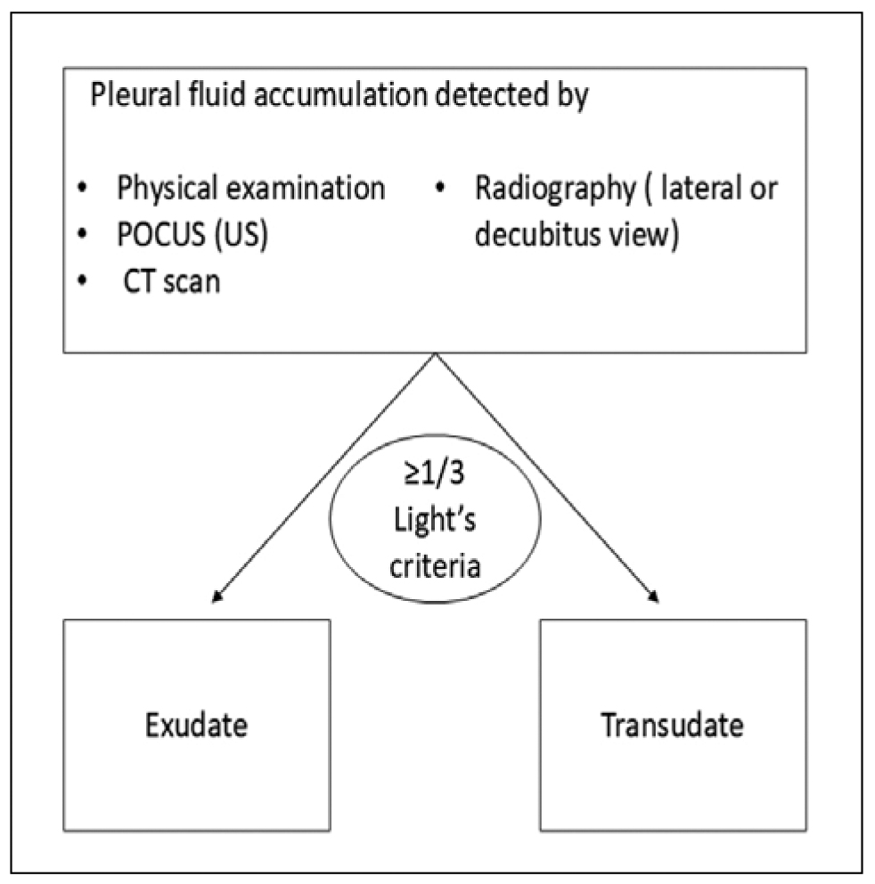
Figure 1. Pleural effusion types
When high suspicion of transudate effusion is present and Light’s criteria indicate an exudate effusion the serum pleural effusion albumin gradient (SPAG) or serum pleural effusion protein gradient (SPPG) can be used as a tool. More specifically SPAG (= serum albumin – pleural albumin) > 1.2g/dl or SPPG (=serum protein – pleural protein) > 3.1 g/dl indicates a transudate effusion [1].
Other potential criteria that can be easily used and show a higher specificity identifying an exudate effusion compared to Light’s are the Heffner’s criteria, which are based on the pleural fluid cholesterol only and without the need for serum values. More specifically, with a cut off value of 45 mg/dl higher values indicate exudate effusions with a reported sensitivity and specificity of 94% and 100%, respectively [25,26].
Another potential way to differentiate exudate from transudate pleural effusions especially in cases where LDH, or protein are not easily available, is the pleural fluid serum bilirubin ratio. Exudates tend to have a greater mean bilirubin ratio compared to transudates. Based on a cross-sectional study in 2018 with a cut-off point of 0.6, the sensitivity, specificity, positive predictive value and negative predictive value were respectively 88.00%, 93.00%, 97.00% & 75.00%, while back in 1990 Meisel et al. revealed the sensitivity of 96% and specificity of 83% [27,28].
Chylothorax
The accumulation of chyle in the pleural space, also known as chylothorax, is a rare but highly morbid cause of exudate pleural effusion. Biochemical analysis of the pleural fluid is necessary for the diagnosis and requires triglyceride levels >110 mg/dl with concomitant cholesterol levels <200 mg/dl, while in cases where levels of triglycerides are less than 55mg/dl the diagnosis is excluded. Based on literature a pleural fluid triglyceride of >110 mg·dL−1 has a 1% chance of being non-chylous and a triglyceride of <50 mg·dL−1 has a 5% chance of being chylous. In borderline cases with high clinical suspicion for chylothorax where pleural fluid triglyceride levels are between 55 and 110mg/dl, such as in cases of malnutrition, the diagnosis can be made with the use of lipoprotein electrophoresis for the detection of chylomicrons in the pleural fluid. As far as the typical gross milky appearance of the fluid is concerned it is seen in only one third of patients. Therefore, the gross appearance is neither sensitive nor specific enough to diagnose a chylothorax, as similar appearances may also be observed in conditions such as pseudochylothorax and pleural infection/empyema. Centrifugation may help in differentiating chylothorax from empyema as in cases of empyema it clears suspended leukocytes and debris leading to a clear supernatant [29–32].
Pathophysiology of Chylothorax
Chyle is transported via the major and minor thoracic duct into systemic circulation. It is composed of interstitial fluid, fluid from the lymphatic system and fluid originating from the intestines which is therefore rich in lymphocytes, immunoglobulins, proteins, lipids, fat soluble vitamins and electrolytes. The loss of this protein-rich, calorie-rich fluid, in cases of chylous leak, can cause serious complications including dehydration, malnutrition and immunosuppression [33].
The anatomical course of the major thoracic duct is complex with considerable variation; therefore, development of chylothorax is dependent on the site and level of the thoracic duct defect.
Causes of chylothorax are mainly traumatic and less frequently non-traumatic. Traumatic chylothorax can be subdivided into iatrogenic, such as after thoracic surgeries or after placement of a central subclavian line or a pacemaker, and non-iatrogenic, caused by external injuries or sudden physical strain. Non-traumatic chylothorax on the other hand, is associated with a wide variety of causes but malignancy remains the major one. Specifically, potential pathologies that directly invade the lymphatic ducts, such as lymphoma and mainly non-Hodgkin lymphoma (in 70% of cases) can cause non-traumatic chylothorax. Another potential mechanism is high hydrostatic pressure inside the lymphatics due to direct obstruction of the thoracic duct or indirectly through increased central venous pressure. Such direct or indirect causes are malignancies, a retrosternal goiter, right heart failure, mediastinal lymph node enlargement, such as in tuberculosis, lymphangioleiomyomatosis, histoplasmosis, sarcoidosis, subclavian vein thrombosis, or superior vena cava obstruction. Interestingly, chylous ascites can also lead to chylothorax through translocation of chyle via pores of the diaphragm. In up to 9% of cases, the etiology remains unknown (termed idiopathic chylothorax) [29,30,34]. However, before jumping to conclusions it is of paramount importance to consider drug-induced chylothorax as part of the differential diagnosis. For example, Chen, Bojiang et al. reported a phenomenically exceptionally rare case of chylothorax induced by Dasatinib, which is a tyrosine kinase inhibitor used for the treatment of BCR-ABL-positive chronic myeloid leukaemia (CML) or Philadelphia chromosome-positive acute lymphoblastic leukaemia (ALL) [35].
Therapeutic approach of chylothorax comprises conservative and invasive choices. The main therapeutic target is to reduce the production and flow of chyle by minimizing long-chain triglyceride (LCT) intake. This type of triglycerides is absorbed by the lymphatic vessels from the interstitial fluid after being converted to chylomicrons within the enterocytes and transported to the systemic circulation via the thoracic duct in the form of chyle.
In order to manage that, a very low-fat diet comprising mainly medium-chain triglycerides (MCT) that consist of saturated fatty acids of 8 to 12 carbon length is preferred. Medium-chain triglycerides are absorbed directly into the portal circulation without micelle formation thus bypassing the lymphatic system and not contributing to chyle production. Another potential solution is the nil-by-mouth approach and the use of parenteral nutrition. The goal is to allow the thoracic duct leak to heal while preventing malnutrition, for a period of 2–4 weeks. While on fat-modified diets, one must take into consideration the potential essential fatty acid deficiency that results when there is insufficient dietary intake of linoleic acid and alpha-linolenic acid from restricted dietary fat consumption. This is because the human body is unable to synthesize linoleic acid and alpha-linolenic acid due to the lack of enzymes that can introduce and nervousdouble bonds beyond the Δ9 site. These essential fatty acids are necessary for healthy cell membrane formation, cholesterol metabolism, blood clotting, as well as proper brain and nervous system development. To prevent this, 2-4% of total calories should arise from linoleic acid and 0.2-1% from alpha-linolenic acid [33,36,37].
Another conservative way to reduce the production of chyle is via the administration of somatostatin analogues (SSAs) like octreotide, or α-adrenergic agonists such as midodrine. Both reduce the production of chyle by the small intestine, reducing its motility and the hepatic and visceral blood flow. Somatostatin analogues should be used when conservative dietary measures fail [36,38].
Regarding invasive therapeutic choices, drainage of the chylous effusion with a chest tube (tube thoracostomy) or catheter may be necessary for symptom relief in parallel with the pharmaceutical approach [30]. In cases where the conservative management fails and for example pleural fluid drainage continues to exceed 1 L/d, surgical thoracic duct ligation or embolization is usually recommended [30].
Pseudochylothorax
Finally, a pathological entity worth mentioning is pseudochylothorax, which can mimic and must be differentiated from chylothorax. It is the result of a chronic persistent exudative pleural effusion that gradually becomes enriched with cholesterol, also termed cholesterol effusion (Table 3). Chronic tuberculous effusion, chronic pneumothorax, rheumatoid effusion, chronic haemothorax and incompletely drained empyema are among common causes of pseudochylothorax. These effusions have the same milky appearance as chylothorax but are differentiated in the pleural fluid analysis since they are characterized by higher levels of cholesterol (>200mg/dl), lower levels of triglycerides (<50mg/dl) and absence of chylomicrons compared to chylothorax [31]. Finally another tool to distinguish between pseudochylothorax and chylothorax, is a fluid to serum cholesterol ratio <1 and triglyceride ratio >1 which is characteristic for chylothorax [39] (Figure 2).
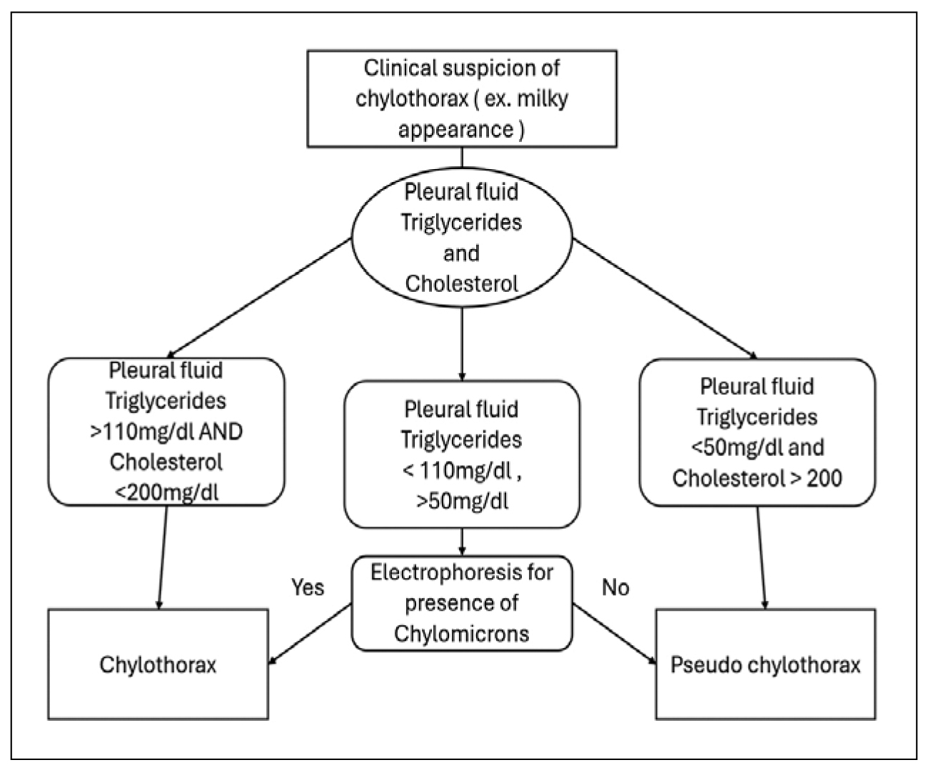
Figure 2. Chylothorax work up.
CONCLUSION
When dealing with a pleural effusion, a well-structured and concise approach should always be the primary goal. A combination of the patient’s history, physical examination, clinical suspicion regarding the potential underlying etiology and use of POCUS to guide thoracocentesis is mandatory to differentiate exudative from transudative effusions and establish a therapeutic approach. Due to the diverse etiological factors and patient characteristics associated with pleural effusion, individualized management strategies are recommended especially in cases of rare entities such as chylothorax and pseudochylothorax where clinical suspicion and a well-established differential diagnosis are of paramount importance for addressing the underlying clinical entity.
Conflict of interest
None to declare.
Declaration of funding
None to declare.
Author contributions
DE conceived the idea; DE, IM, PK performed the literature search; DE wrote the manuscript; ML critically revised the manuscript; ML oversaw the study, DE, IM, PK, ML revised the manuscript.
REFERENCES
- D’Agostino HP, Edens MA. Physiology, Pleural Fluid. [Updated 2023 Aug 28]. In: StatPearls [Internet]. Treasure Island (FL): StatPearls Publishing; 2026 Jan-. Available from: https://www.ncbi.nlm.nih.gov/books/NBK513353/
- Krishna R, Antoine MH, Alahmadi MH, Rudrappa M. Pleural Effusion. [Updated 2024 Aug 31]. In: StatPearls [Internet]. Treasure Island (FL): StatPearls Publishing; 2026 Jan-. Available from: https://www.ncbi.nlm.nih.gov/books/NBK448189/
- Feller-Kopman D, Light R. Pleural Disease. N Engl J Med. 2018;378(8):740–51. doi:10.1056/NEJMra1403503
- Ilahi M, St Lucia K, Ilahi TB. Anatomy, Thorax, Thoracic Duct. [Updated 2023 Jul 24]. In: StatPearls [Internet]. Treasure Island (FL): StatPearls Publishing; 2026 Jan-. Available from: https://www.ncbi.nlm.nih.gov/books/NBK513227/.
- DeBiasi EM, Feller-Kopman D. Anatomy and Applied Physiology of the Pleural Space. Clin Chest Med. 2021;42(4):567–76. doi:10.1016/j.ccm.2021.08.005
- Iyer NP, Reddy CB, Wahidi MM, Lewis SZ, Diekemper RL, Feller-Kopman D, et al. Indwelling Pleural Catheter versus Pleurodesis for Malignant Pleural Effusions. A Systematic Review and Meta-Analysis. Ann Am Thorac Soc. 2019;16(1):124–31. doi:10.1513/AnnalsATS.201807-495OC
- Muruganandan S, Azzopardi M, Thomas R, Fitzgerald DB, Kuok YJ, Cheah HM, et al. The Pleural Effusion And Symptom Evaluation (PLEASE) study of breathlessness in patients with a symptomatic pleural effusion. Eur Respir J. 2020;55(5):1900980. doi:10.1183/13993003.00980-2019
- Estenne M, Yernault JC, De Troyer A. Mechanism of relief of dyspnea after thoracocentesis in patients with large pleural effusions. Am J Med. 1983;74(5):813–9. doi:10.1016/0002-9343(83)91072-0
- Wong CL, Holroyd-Leduc J, Straus SE. Does This Patient Have a Pleural Effusion? JAMA. 2009;301(3):309-17. doi:10.1001/jama.2008.937
- Karkhanis V, Joshi J. Pleural effusion: diagnosis, treatment, and management. Open Access Emerg Med. 2012;4:31-52. doi:10.2147/OAEM.S29942
- Yousefifard M, Baikpour M, Ghelichkhani P, Asady H, Shahsavari Nia K, Moghadas Jafari A, et al. Screening Performance Characteristic of Ultrasonography and Radiography in Detection of Pleural Effusion; a Meta-Analysis. Emerg (Tehran). 2016;4(1):1–10.
- Zaki HA, Albaroudi B, Shaban EE, Shaban A, Elgassim M, Almarri ND, et al. Advancement in pleura effusion diagnosis: a systematic review and meta-analysis of point-of-care ultrasound versus radiographic thoracic imaging. Ultrasound J. 2024;16(1):3. doi:10.1186/s13089-023-00356-z
- Sikora K, Perera P, Mailhot T, Mandavia D. Ultrasound for the Detection of Pleural Effusions and Guidance of the Thoracentesis Procedure. ISRN Emerg Med. 2012;2012(1):1–10. doi:10.5402/2012/676524
- Zhang Y, Zhang Y, Wang W, Feng X, Guo J, Chen B, et al. Diagnostic accuracy of thoracic CT to differentiate transudative from exudative pleural effusion prior to thoracentesis. Respir Res. 2024;25(1):53. doi:10.1186/s12931-024-02681-w
- Wiederhold BD, Sharma S, O’Rourke MC. Thoracentesis. [Updated 2024 Oct 6]. In: StatPearls [Internet]. Treasure Island (FL): StatPearls Publishing; 2026 Jan-. Available from: https://www.ncbi.nlm.nih.gov/books/NBK441866/
- Shen-Wagner J, Gamble C, MacGilvray P. Pleural Effusion: Diagnostic Approach in Adults. Am Fam Physician. 2023;108(5):464–75.
- Light RW. A New Classification of Parapneumonic Effusions and Empyema. Chest. 1995;108(2):299–301. doi:10.1378/chest.108.2.299
- Colice GL, Curtis A, Deslauriers J, Heffner J, Light R, Littenberg B, et al. Medical and Surgical Treatment of Parapneumonic Effusions. Chest. 2000;118(4):1158–71. doi:10.1378/chest.118.4.1158
- Wu H, Khosla R, Rohatgi PK, Chauhan SS, Paal E, Chen W. The minimum volume of pleural fluid required to diagnose malignant pleural effusion: A retrospective study. Lung India. 2017;34(1):34–7. doi:10.4103/0970-2113.197120
- Porcel JM, Vives M, Cao G, Esquerda A, Rubio M, Rivas MC. Measurement of pro-brain natriuretic peptide in pleural fluid for the diagnosis of pleural effusions due to heart failure. Am J Med. 2004;116(6):417–20. doi:10.1016/j.amjmed.2003.11.008
- Jany B, Welte T. Pleural Effusion in Adults—Etiology, Diagnosis, and Treatment. Dtsch Arztebl Int. 2019;116(21):377-86. doi:10.3238/arztebl.2019.0377
- Antony VB. DRUG-INDUCED PLEURAL DISEASE. Clin Chest Med. 1998;19(2):331–40. doi:10.1016/S0272-5231(05)70080-0
- Sharma K, Fultariya L, Reddy Mallimala P, Shah K, Sharma V. Comparison of the Efficacy of Light’s Criteria With Serum-Effusion Albumin Gradient and Pleural Effusion Glucose. Cureus. 2023;15(8):e43319. doi:10.7759/cureus.43319
- Romero-Candeira S, Fernández C, Martı́n C, Sánchez-Paya J, Hernández L. Influence of diuretics on the concentration of proteins and other components of pleural transudates in patients with heart failure. Am J Med. 2001;110(9):681–6. doi:10.1016/S0002-9343(01)00726-4
- Devkota KC, Hamal S, Panta P. Comparison of Heffner Criteria and Light Criteria in Differentiating Exudative and Transudative Pleural Effusion. Nepal Med Coll J. 2020;22(3):141–5. doi:10.3126/nmcj.v22i3.32638
- Gautam S, K C SR, Bhattarai B, K C G, Adhikari G, Gyawali P, et al. Diagnostic value of pleural cholesterol in differentiating exudative and transudative pleural effusion. Ann Med Surg (Lond). 2022;82:104479. doi:10.1016/j.amsu.2022.104479
- Agrawal P, Shrestha TM, Prasad PN, Aacharya RP, Gupta P. Pleural Fluid Serum Bilirubin Ratio for Differentiating Exudative and Transudative Effusions. JNMA J Nepal Med Assoc. 2018;56(211):662–5.
- Meisel S, Shamiss A, Thaler M, Nussinovitch N, Rosenthal T. Pleural Fluid to Serum Bilirubin Concentration Ratio for the Separation of Transudates from Exudates. Chest. 1990;98(1):141–4. doi:10.1378/chest.98.1.141
- Ur Rehman K, Sivakumar P. Non-traumatic chylothorax: diagnostic and therapeutic strategies. Breathe. 2022;18(2):210163. doi:10.1183/20734735.0163-2021
- Bhatnagar M, Fisher A, Ramsaroop S, Carter A, Pippard B. Chylothorax: pathophysiology, diagnosis, and management—a comprehensive review. J Thorac Dis. 2024;16(2):1645–61. doi:10.21037/jtd-23-1636
- McGrath EE, Blades Z, Anderson PB. Chylothorax: Aetiology, diagnosis and therapeutic options. Respir Med. 2010;104(1):1–8. doi:10.1016/j.rmed.2009.08.010
- Maldonado F, Hawkins FJ, Daniels CE, Doerr CH, Decker PA, Ryu JH. Pleural Fluid Characteristics of Chylothorax. Mayo Clin Proc. 2009;84(2):129–33. doi:10.4065/84.2.129
- Bibby AC, Maskell NA. Nutritional management in chyle leaks and chylous effusions. Br J Community Nurs. 2014;19(Suppl 11):S6–8. doi:10.12968/bjcn.2014.19.Sup11.S6.
- Gary P, Gianniosis M, Ryu J, Flynn S. Chylothorax as a complication of Waldenström macroglobulinaemia with a patient’s perspective. BMJ Case Rep. 2021;14(5):e241707. doi:10.1136/bcr-2021-241707
- Chen B, Wu Z, Wang Q, Li W, Cheng D. Dasatinib-induced chylothorax: report of a case and review of the literature. Invest New Drugs. 2020;38(5):1627–32. doi:10.1007/s10637-020-00932-3
- Fogg KL, Trauth A, Horsley M, Vichayavilas P, Winder M, Bailly DK, et al. Nutritional management of postoperative chylothorax in children with CHD. Cardiol Young. 2023;33(9):1663–71. doi:10.1017/S1047951122003109
- Agrawal A, Chaddha U, Shojaee S, Nadolski G, Liberman M, Lee YCG, et al. Multidisciplinary management of adult patients with chylothorax: a consensus statement. Eur Respir J. 2024;64(5):2400470. doi:10.1183/13993003.00470-2024
- Sharkey AJ, Rao JN. The successful use of octreotide in the treatment of traumatic chylothorax. Tex Heart Inst J. 2012;39(3):428–30.
- Nair SK, Petko M, Hayward MP. Aetiology and management of chylothorax in adults. Eur J Cardiothorac Surg. 2007;32(2):362–9. doi:10.1016/j.ejcts.2007.04.024.